KOSIN UNIVERSITY COLLEGE OF MEDICINE
Articles
- Page Path
- HOME > Kosin Med J > Volume 33(1); 2018 > Article
-
Case Report
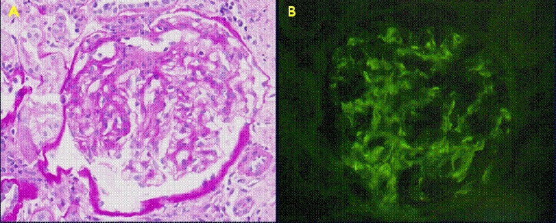
IgA nephropathy in a patient with ankylosing spondylitis well controlled with etanercept - Do-Hyeong Lee, Geun-Tae Kim, Na-Kyoung Hwang, Eun-Heui Kim
-
Kosin Medical Journal 2018;33(1):85-90.
DOI: https://doi.org/10.7180/kmj.2018.33.1.85
Published online: June 30, 2018
1Division of Rheumatology, Department of Internal Medicine, Kosin University College of Medicine, Busan, Korea.
2Division of Rheumatology, Department of Internal Medicine, Pusan National University Hospital, Busan, Korea.
- Corresponding Author: Geun-Tae Kim, Division of Rheumatology, Department of Internal Medicine, College of Medicine, Kosin University, 262, Gamchen-ro, Seo-gu, Busan 49267, Korea. Tel: +82-51-990-6154, Fax: +82-51-990-3010, gtah@hanmail.net
• Received: July 9, 2015 • Revised: August 21, 2015 • Accepted: September 3, 2015
Copyright © 2018 Kosin University College of Medicine
- 1,227 Views
- 5 Download
- 1 Crossref
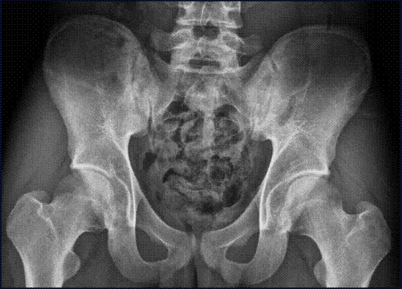

Figure & Data
References
Citations
Citations to this article as recorded by 
- IgA Nephropathy—Ankylosing Spondylitis–Associated or Adalimumab-Induced?
Joana Ricardo Pires, Anabela Tavares Valadão Barcelos
JCR: Journal of Clinical Rheumatology.2021; 27(8S): S449. CrossRef
 PubReader
 Cite
Cite