KOSIN UNIVERSITY COLLEGE OF MEDICINE
Articles
- Page Path
- HOME > Kosin Med J > Volume 28(2); 2013 > Article
-
Original Article
Clinical Features and Histological Findings of 17 Patients with Chronic Actinic Dermatitis - Min Soo Jang, Kang Hoon Lee, Sang Hwa Han, Jong Bin Park, Dong Young Kang, Sang Tae Kim, Kee Suck Suh
-
Kosin Medical Journal 2013;28(2):145-153.
DOI: https://doi.org/10.7180/kmj.2013.28.2.145
Published online: January 19, 2013
Department of Dermatology, College of Medicine, Kosin University, Busan, Korea
- Corresponding author: Kee Suck Suh, Department of Dermatology, College of Medicine, Kosin University, 34 Amnamdong, Seo-gu, Busan, 602-702, Korea TEL: +82-51-990-6145 FAX: +82-51-990-3041 E-mail: ksderm98@unitel.co.kr
• Received: July 18, 2013 • Accepted: September 24, 2013
Copyright © 2013 Kosin University School of Medicine Proceedings
This is an Open Access article distributed under the terms of the Creative Commons Attribution Non-Commercial License (http://creativecommons.org/licenses/by-nc/3.0/) which permits unrestricted non-commercial use, distribution, and reproduction in any medium, provided the original work is properly cited.
- 863 Views
- 2 Download
Abstract
-
Objectives
- This study was designed to evaluate the clinical, histological and immunohistochemical findings and treatment of chronic actinic dermatitis in Korean patients.
-
Methods
- Seventeen Korean patients with chronic actinic dermatitis were enrolled for this study. The clinical and histological findings and the results of phototest were reviewed with medical records, clinical photographs and pathologic slides. We also reviewed the effectiveness of the treatments in all patients with chronic actinic dermatitis.
-
Results
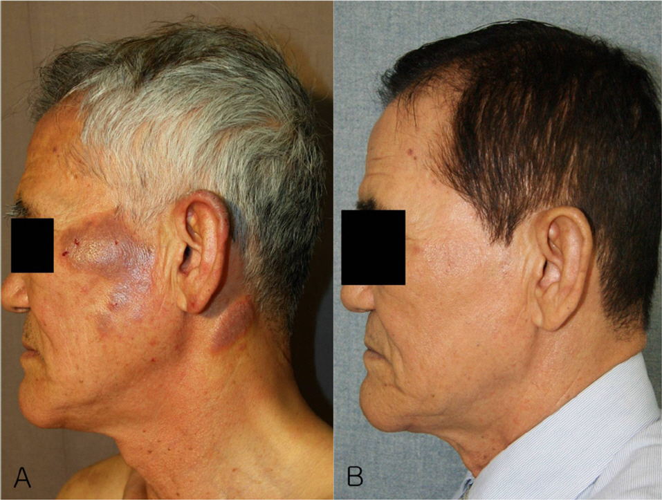
- In all patients with chronic actinic dermatitis, pruritus was severe, and the patients present in the early stages with erythemas on the face, neck and the back of the hands. As the eruption progresses, it became lichenified and scaly plaques and papules developed. The face, upper extremity and neck were most commonly affected. The most common abnormal results of the phototests were decreased MED-UVB alone. In 8 patients with actinic reticuloid, histopathologic findings showed irregular acanthosis, parakeratosis, spongiosis, atypical hyperchromatic cells with cerebriform nuclei, epidermotropism, Pautrier-like microabscess, deep perivascular lymphocytic infiltrates, vertically-streaked collagen in the papillary dermis, stellate and multinucleated fibroblasts. Treatment includes topical tacrolimus and corticosteroid, oral corticosteroid, azathioprine and cyclosporine.
-
Conclusions
- Our study showed classic clinical and histological findings. The most common abnormal results of the phototests were decreased MED-UVB alone. Topical steroid, tacrolimus and systemic cyclosporine, azathioprine are effective in treating chronic actinic dermatitis.
Fig. 1.(A) Irregular acanthosis, and lymphocytic infiltration in the papillary dermis and reticular dermis (H&E, x 40), (B) Epidermotropism, irregular acanthosis and a perivascular infiltrate of the papillary dermis (H&E, x 100) (Case 16).
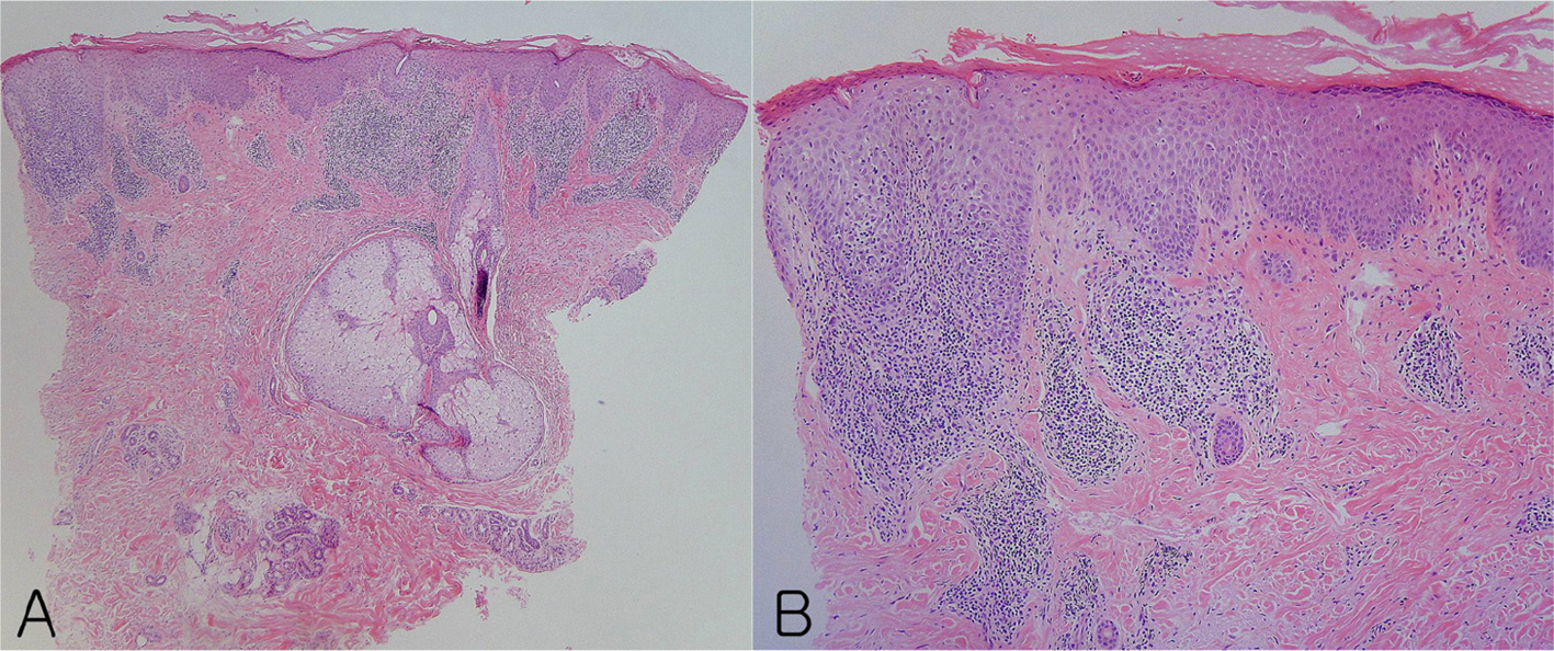
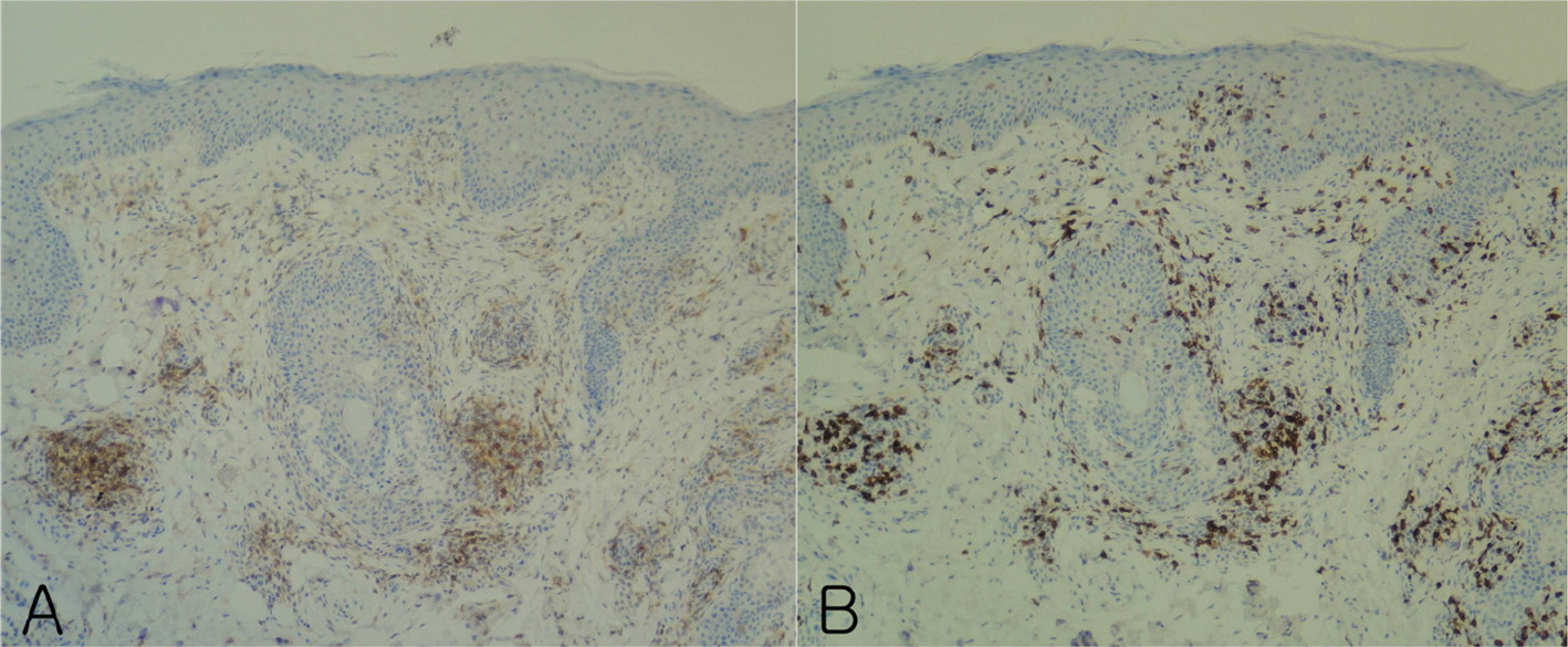
Fig. 2.Immunohistochemical studies showed that CD4 positive T cells (A) and CD8 positive T cells (B) were in the epidermis and dermis (x 100). CD8 positive T cells predominated in the epidermis (B) compared to CD4 positive T cells (A). (Case 6).

Fig. 3.Immunohistochemical studies showed that CD4 positive T cells (A) and CD8 positive T cells (B) were in the epidermis and dermis (x 100). CD8 positive T cells predominated in the epidermis (B) compared to CD4 positive T cells (A). (Case 6).

Fig. 4.Immunohistochemical studies showed that CD4 positive T cells (A) and CD8 positive T cells (B) were in the epidermis and dermis (x 100). CD8 positive T cells predominated in the epidermis (B) compared to CD4 positive T cells (A). (Case 6).
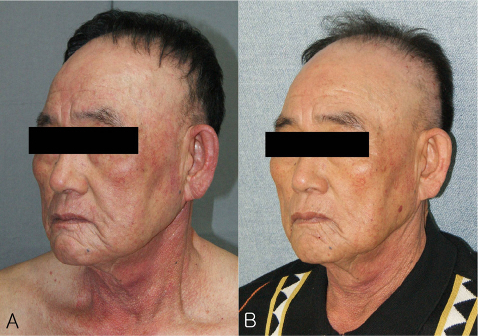
Table 1.Summary of clinical characteristics in patients with chronic actinic dermatitis
Table 2.Results of phototests in patients with chronic actinic dermatitis
Table 3.Results of photopatch tests in patients with chronic actinic dermatitis
| Case | 1 | 2 | 3 | 4 | 5 | 6 | 7 | 8 | 9 | 10 | 11 | 12 | 13 | 14 | 15 | 16 | 17 |
|---|---|---|---|---|---|---|---|---|---|---|---|---|---|---|---|---|---|
|
Photo patch test |
- | DN | DN | - | - | - | DN | - | DN | - | DN | - | DN | DN | DN | DN | DN |
Table 4.Summary of treatment in patients with chronic actinic dermatitis
- 1.Hawk JL, Lim HWChronic actinic dermatitis. In: Lim HW, Hönigsmann H, Hawk JLeditors.., editors. Photodermatology. 1st ed.New York: Informa Healthcare; 2007. p. 169–83.Article
- 2.Hawk JL. Chronic actinic dermatitis. Photodermatol Pho-toimmunol Photomed 2004;20:312–4.Article
- 3.Uetsu N, Okamoto H, Fujii K, Doi R, Horio T. Treatment of chronic actinic dermatitis with tacrolimus ointment. J Am Acad Dermatol 2002;47:881–4.ArticlePubMed
- 4.Ma Y, Lu Z. Treatment with topical tacrolimus favors chronic actinic dermatitis: a clinical and immunopathological study. J Dermatolog Treat 2010;21:171–7.ArticlePubMed
- 5.Cho HJ, Hann SK, Shin HK, Park YK, Lee KH. Effects and significance of cyclosporine therapy in chronic actinic dermatitis. Korean J Dermatol 1997;35:458–64.
- 6.Dawe RS, Ferguson J. Diagnosis and treatment of chronic actinic dermatitis. Dermatol Ther 2003;16:45–51.ArticlePubMed
- 7.Stinco G, Codutti R, Frattasio A, De Francesco V, Patrone P. Chronic actinic dermatitis treated with cyclosporine-A. Eur J Dermatol 2002;12:455–7.PubMed
- 8.Kim JK, Li Chen, Kim TH. Three cases of chronic actinic dermatitis treated with systemic PUVA therapy. Ann Dermatol 1997;9:197–200.Article
- 9.Beach RA, Pratt MD. Chronic actinic dermatitis: clinical cases, diagnostic workup, and therapeutic management. J Cutan Med Surg 2009;13:121–8.ArticlePubMed
- 10.Bakels V, van Oostveen JW, Preesman AH, Meijer CJ, Willemze R. Differentiation between actinic reticuloid and cutaneous T cell lymphoma by T cell receptor gamma gene rearrangement analysis and immunophenotyping. J Clin Pathol 1998;51:154–8.ArticlePubMedPMC
- 11.Heller P, Wieczorek R, Waldo E, Meola T, Buchness MR, Soter NA, et al. Chronic actinic dermatitis: An immunohistochemical study of its T-cell antigenic profile, with comparison to cutaneous T-cell lymphoma. Am J Dermatopathol 1994;16:510–6.PubMed
- 12.Toonstra J, van der Putte SC, van Wichen DF, van Weelden H, Henquet CJ, van Vloten W. Actinic reticuloid: immunohistochemical analysis of the cutaneous infiltrate in 13 patients. Br J Dermatol 1989;120:779–86.ArticlePubMed
- 13.Kim KH, Nam JT, Joh GY. A case of chronic actinic dermatitis. Korean J Dermatol 1992;30:906–12.
- 14.Lim HW, Morison WL, Kamide R, Buchness MR, Harris R, Soter NA. Chronic actinic dermatitis: An analysis of 51 patients evaluated in the United States and Japan. Arch Dermatol 1994;130:1284–9.ArticlePubMed
- 15.Won CH, Youn JI. Clinical study of 12 cases with chronic actinic dermatitis. Korean J Dermatol 2007;45:1144–8.
- 16.Sim SJ, Eim JJ, Song KH, Joh GY, Kim KH. Results of photopatch and patch tests in 35 Korean patients with chronic actinic dermatitis and clinical importance of these findings. Korean J Dermatol 2004;42:976–82.
- 17.Yap LM, Foley P, Crouch R, Baker C. Chronic actinic dermatitis: a retrospective analysis of 44 cases referred to an Australian photobiology clinic. Australas J Dermatol 2003;44:256–62.ArticlePubMed
- 18.Tan AW, Lim KS, Theng C, Chong WS. Chronic actinic dermatitis in Asian skin: a Singaporean experience. Photodermatol Photoimmunol Photomed 2011;27:172–5.ArticlePubMed
- 19.Jeon YS, Suh KS, Kim ST. A comparison of minimal erythema dose and minimal melanogenic dose induced by ultraviolet A-1, broad band ultraviolet A, narrow band ultraviolet B, and broad band ultraviolet B. Korean J Dermatol 2005;43:442–9.
- 20.Hawk JL, Magnus IA. Chronic actinic dermatitis: an idiopathic photosensitivity syndrome including actinic reticuloid and photosensitive eczema. Br J Dermatol 1979;101:24.PubMed
- 21.Norris PG, Hawk JL. Chronic actinic dermatitis: a unifying concept. Arch Dermatol 1990;126:376–8.ArticlePubMed
- 22.Ive FA, Magnus IA, Warin RP, Jones EW. "Actinic reticuloid"; a chronic dermatosis associated with severe photosensitivity and the histological resemblance to lymphoma. Br J Dermatol 1969;81:469–85.ArticlePubMed
- 23.Zak-Prelich M, Schwartz RA. Actinic reticuloid. Int J Dermatol 1999;38:335–42.ArticlePubMed
- 24.Chew AL, Bashir SJ, Hawk JL, Palmer R, White IR, McFadden JP. Contact and photocontact sensitization in chronic actinic dermatitis: a changing picture. Contact Dermatitis 2010;62:42–6.ArticlePubMed
- 25.Que SK, Brauer JA, Soter NA, Cohen DE. Chronic actinic dermatitis: an analysis at a single institution over 25 years. Dermatitis 2011;22:147–54.ArticlePubMed
- 26.Kim YC, Song WK. A comparison of UVB - induced minimal erythema dose (MED)s to the skin of the back and extremities in young adult Koreans. Korean J Dermatol 1998;36:261–5.
- 27.De Silva BD, McLaren K, Kavanagh GM. Photosensitive mycosis fungoides or actinic reticuloid? Br J Dermatol 2000;142:1221–7.ArticlePubMed
- 28.Cerroni L, Gatter K, Kerl HSkin lymphoma: the illustrated guide. 3rd ed.Oxford: Wiley-Black well; 2009. p. 11–56.
- 29.Goodlad J, Calonje ECutaneous lymphoproliferative diseases and related disorders. In: Calonje E, Brenn T, Lazar A, McKee PHeditors.., editors. McKee's pathology of the skin with clinical correlations. 4th ed.Philadelphia: Elsevier; 2011. p. 1311–420.
- 30.Weedon DWeedon's skin pathology. 3rd ed.London: Churchill Livingstone Elsevier; 2010. p. 525–40.
References
Figure & Data
References
Citations
Citations to this article as recorded by 
 PubReader
 ePub Link
 Cite
Cite