 KOSIN UNIVERSITY COLLEGE OF MEDICINE
Articles
- Page Path
- HOME > Kosin Med J > Volume 33(3); 2018 > Article
-
Case Report
A Case of Spontaneous Common Iliac Atery Dissection - Juho Noh, Il Rhee, Minsung Kim, Jonghyun Lee, Kisu Kim, Byungwhan Park
-
Kosin Medical Journal 2018;33(3):431-437.
DOI: https://doi.org/10.7180/kmj.2018.33.3.431
Published online: December 31, 2018
Division of Cardiology, Department of Internal Medicine, Good Samsun Hospital, Busan, Korea.
- Corresponding Author: Il Rhee, Department of Internal Medicine, Good Samsun Hospital, 326 Gaya-daero, Sasang-gu, Busan, 47007, Korea. Tel: +82-51-322-0900, Fax: +82-51-323-3308, cormed21@gmail.com
• Received: July 29, 2016 • Revised: September 21, 2016 • Accepted: October 12, 2016
Copyright © 2018 Kosin University College of Medicine
Articles published in Kosin Medical Journal are open-access, distributed under the terms of the Creative Commons Attribution Non-Commercial License (http://creativecommons.org/licenses/by-nc/4.0/) which permits unrestricted non-commercial use, distribution, and reproduction in any medium, provided the original work is properly cited.
- 1,393 Views
- 3 Download
- 1 Crossref
Abstract
-
- Spontaneous and isolated dissection of the limb arteries without involvement of the aorta is extremely rare, and has been reported previously in pregnant patients in association with collagen vascular disease, and in cases of high-energy trauma or intensive activity in athletes. There is no consensus yet on indications for medical or surgical therapeutic modality. Due to the rarity of spontaneous dissection of external iliac artery, its natural history has been poorly described.
- A healthy 50-year-old male with normotension was admitted with an acute onset of left flank pain. Left external iliac artery dissection was diagnosed by abdominal computed tomography.
- A 50-year-old man visited the urology department of the authors' hospital with left back pain that started two weeks ago. The patient had smoked for 30 years. He had no unusual history of cardiovascular diseases and connective tissue diseases, with the exception of having undergone excision of a lipoma of the left lower scapula seven years ago. He had no unusual family history, either. At the time of the visit, the patient had acute pain and complained of left flank pain. His vital signs were as follows: blood pressure 110/70 mmHg, pulse 120 times/minute, respiratory rate 20 times/minute and body temperature 36.2 ℃. When auscultated on the chest, the patient had normal pulmonary and cardiac sounds while when palpitated on the abdomen, he complained of tenderness over the left upper abdomen and left costovertebral angle. No specific findings were observed in the bilateral lower extremities.
- At the time of the visit, the patient's peripheral blood test results were as follows: leukocyte 11,070/mm3 (neutrophil 57.6%, lymphocyte 32.3%, eosinophil 1.05%), hemoglobin 13.2g/dL and platelet 241,000/mm3. The serum biochemical test results were as follows: albumin 3.9 g/dl, total bilirubin 0.19 mg/dL, direct bilirubin 0.06 mg/dl, AST 13 IU/L, ALT 11 IU/L, ALP 262 IU/L, prothrombin time 10.5 sec, PT INR 0.99 (normal range: 0.85–1.15), total cholesterol 177 mg/dl, HDL 32 mg/dl, LDL 97 mg/dl, TG 533 mg/dl, Troponin I 0.1 ng/ml (normal range: 0–0.3), CK-MB 0.83 ng/ml (normal range: 0–4.87), D-dimer 545.77 ng/ml (normal range: 0–500), blood urea nitrogen 19.2 mg/dl, creatinine 1.8 mg/dl, uric acid 4.2 mg/dl, LDL 264 and CRP 0.65 mg/dl. Other serum electrolyte and urine tests showed no specific findings and α-1-Antitrypsin and ANCA (anti-neutrophil cytoplasmic antibody) were not checked.
- There was no evidence of urinary stone in a simple urinary tract x-ray. An abdominal computed tomography (CT) scan revealed a mass lesion over the upper left psoas muscle and left hydronephrosis. So a contrast-enhanced CT scan was also performed. As a result, features of dissection on the left external iliac artery origin accompanying a hematoma and left hydronephrosis due to the compression of the hematoma were found (Fig. 1); therefore, this case was transferred to the cardiopulmonary department, where an ABI (ankle brachial index) test and transthoracic echocardiography were additionally performed. The findings were normal.
- The authors diagnosed the patient as spontaneous left external iliac artery dissection and decided to perform PTA (Percutaneous transluminal angioplasty). The left femoral artery was punctured and a 12 French catheter was inserted, followed by a 0.035-inch guidewire (Terum, Tokyo, Japan). With the performance of external iliac artery angiography, a graft stent (W. L GORE VIABAHN®, Flagstaff, AZ, USA) with a diameter of 11 mm and a length of 50 mm was placed on the dissected vessel site (Fig. 2A). To confirm a proper dissection sealing, imaging of the vessel using IVUS(Intravascular ultrasound) was conducted and it showed no abnormal findings (Fig. 2B). Afterwards, external iliac artery angiography was performed again to confirm that the vessel was well visualized to the distal part, with which the procedure was terminated (Fig. 3).
- After the stent implantation, the patient showed an alleviation of the left lower abdomen pain and left flank pain. A week later, an abdominal CT scan revealed the disappearance of the left iliac artery dissection; however, there was no improvement in the hydronephrosis due to the hematoma around the dissected site and the ureteral compression on the left distal part. Serum creatinine level did not improve, either, so the treatment was commissioned to the urology department where a double J catheter insertion was performed; after this, the patient was discharged.
- The abdominal CT scan performed a month later confirmed the disappearance of the hematoma and hydronephrosis near the left external iliac artery and a decrease in serum creatinine level (1.34 mg/dl). So the D-J catheter was removed, following which the patient has been monitored in the outpatient clinic.
CASE
- Spontaneous external iliac artery dissection is a very rare disease and it is known to be caused by trauma, connective tissue diseases, fibrous dysplasia, lack of alpha 1-trypsin, vascular ulcer due to atherosclerosis, persistent and strong external force and so on. According to some reports published in the past, pain in the groin area, radicular pain in the back or thigh and lower extremity claudication are the main symptoms.234 Therefore, it can be predicted that its diagnosis will take a long time in many cases. This patient also complained of a sudden onset of left flank pain, and the examinations to detect urologic problems such as urinary stones preceded the diagnosis. Ultrasonography or contrast-enhanced CT may be performed for the diagnosis of the diseases, but vascular angiographic imaging is necessary for the identification of the degree of dissection and the location of intimal flaps.
- In this case experienced by the authors, the patient was a healthy 50-year-old man who had no risk factors for atherosclerosis other than smoking and had no suspected connective tissue diseases or vascular diseases. He had engaged in extended physical exercise such as golf about a month before visiting the authors' hospital, but it was difficult to define this as a clear predisposing factor. Kevin D. et al. presumed, in their case report, that long-term smoking can cause vascular endothelial cell damage, atherosclerosis and aneurysm expansion in the blood vessels that have been relatively largely damaged by intense exercise such as running and swimming, leading to spontaneous iliac artery dissection.5 However, this case showed no evidence of atherosclerosis or aneurysm expansion as in the patient of the case reported by Kevin D. et al. in the abdominal and lower limb arterial angiography.
- Percutaneous intervention, drug treatment and surgical treatment are all possible for lower extremity vascular dissection including external iliac artery dissection.4567 However, in cases where patients have severe symptoms or a high risk of rupture, there is a need for an immediate treatment. According to Whchulis A. R. et al., early surgical treatment is recommended because of a high probability of rupture within months of iliac artery dissection.8 In addition, Honjo O. et al. reported that 8 of 10 patients with iliac artery dissection underwent artificial blood vessel or autologous vein transplantation, which resulted in a successful treatment without a need for subsequent reoperation.9
- Endovascular Tx. has the merit of being relatively easy and noninvasive compared to surgical Tx. but there has been no case of long-term monitoring so far. Kwak, Han et al. reported that percutaneous stenting was performed in two cases of spontaneous iliac artery dissection and there was no recurrence or stent stenosis during a 2 year and 10 month monitoring period.10
- Since the first successful coronary artery intervention in 1977, percutaneous intervention for peripheral arteries as well as for coronary arteries have seen remarkable progresses. The American Heart Association presented recommendations for the treatment of leg arterial intervention in 2005,11 and the revised version was published in 2011. In Europe and North America as well, the Transatlantic Intersociety Consensus Working Group was formed in 2001, centered on the departments of interventional radiology, vascular surgery and cardiology. This group has continuously published and revised treatment guidelines until recently.12 However, these guidelines mostly include content related to the stenosis and occlusion of lower limb arteries associated with arteriosclerosis or diabetes. The authors successfully performed stent implantation using percutaneous intervention in the patient with spontaneous iliac artery dissection, and confirmed that there were no specific problems such as recurrence or restenosis, although the monitoring was only carried out for a relatively short period of seven months. Based on this, the authors believe that objective and systematic guidelines for the dissection and rupture of peripheral arteries including iliac arteries as reported in this case are necessary.
DISCUSSION
- 1. Guthrie W, Maclean H. Dissecting aneurysms of arteries other than the aorta. J Pathol 1972;108:219–235.ArticlePubMed
- 2. The LG, Sieunarine K, Van Schie G, Vasudevan T. Spontaneous common iliac artery dissection after exercise. J Endovasc Ther 2003;10:163–166.ArticlePubMed
- 3. Willson TD, Revese E, Podbielski FJ, Blecha MJ. External iliac artery dissection secondary to endofibrosis in a cyclist. J Vasc Surg 2010;52:219–221.ArticlePubMed
- 4. Savolainen H, Heller G, Fleiscmann A, Widmer MK, Carrel TP, Schmidli J. Spontaneous dissection of common iliac artery. A case report. Vasc Endovascular Surg 2004;38:263–265.ArticlePubMedPDF
- 5. Goodman KD, Sule HP. Bilateral spontaneous dissecting common iliac artery aneurysms: a rare presentation. J Emerg Med 2013;44:757–759.ArticlePubMed
- 6. Lück I, Hanschke D, Geissler C, Gruss JD. [Spontaneous dissection of the external iliac artery due to fibromuscular dysplasia]. Vasa 2003;31:115–121.Article
- 7. Fernández AL, Herreros JM. Spontaneous and isolated dissection of the common iliac artery. J Cardiovasc Surg (Torino) 1997;38:377–379.PubMed
- 8. Whchulis AR, Kincaid OW, Wallace RB. Primary dissecting aneurysm of peripheral arteries. Mayo Clin Proc 1969;44:804–810.PubMed
- 9. Honjo O, Yamada Y, Kuroko Y, Kushida Y, Une D, Hioki K. spontaneous dissection and rupture of common iliac artery in patient with fibromuscular dysplasia: a case report and review of the literature on iliac artery dissections secondary to fibromuscular dysplasia. J Vasc Surg 2004;40:1032–1036.ArticlePubMed
- 10. Kwak HS, Han YM, Chung GH, Yu HC, Jeong YJ. Isolated spontaneous dissection of the common iliac artery: percutaneous stent placement in two patients. Cardiovasc Intervent Radiol 2006;29:883–885.ArticlePubMed
- 11. Hirsch AT, Haskal ZJ, Hertzer NR, et al. ACC/AHA 2005 Practice Guidelines for the management of patients with peripheral arterial disease (lower extremity, renal, mesenteric, and abdominal aortic): a collaborative report from the American Association for Vascular Surgery/Society for Vascular Surgery, Society for Cardiovascular Angiography and Interventions, Society for Vascular Medicine and Biology, Society of Interventional Radiology, and the ACC/AHA Task Force on Practice Guidelines (Writing Committee to Develop Guidelines for the Management of Patients With Peripheral Arterial Disease): endorsed by the American Association of Cardiovascular and Pulmonary Rehabilitation; National Heart, Lung, and Blood Institute; Society for Vascular Nursing; TransAtlantic Inter-Society Consensus; and Vascular Disease Foundation. Circulation 2006;113:e463–e654.PubMed
- 12. Tendera M, Aboyans V, Bartelink ML, et al. ESC Guidelines on the diagnosis and treatment of peripheral artery diseases Document covering atherosclerotic disease of extracranial carotid and vertebral, mesenteric, renal, upper and lower extremity arteries The Task Force on the Diagnosis and Treatment of Peripheral Artery Diseases of the European Society of Cardiology (ESC). Eur Heart J 2011;32:2851–2906.ArticlePubMed
References
Fig. 1
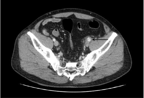
Contrast-enhanced abdominal CT scan showing an intimal flap involving the left external iliac artery with contrast filling of the false and true lumens with surrounding hematoma formation (arrows).
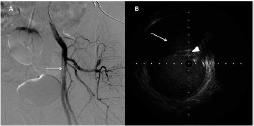
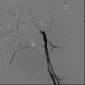
Figure & Data
References
Citations
Citations to this article as recorded by 
- Management of a spontaneous dissection of the right external iliac artery
S. El Hadhri, M. Gueldich, H. Ben Jemaa, I. Frikha
JMV-Journal de Médecine Vasculaire.2021; 46(5-6): 246. CrossRef
 PubReader
 Cite
Cite


