 KOSIN UNIVERSITY COLLEGE OF MEDICINE
Articles
- Page Path
- HOME > Kosin Med J > Volume 32(2); 2017 > Article
-
Case Report
Spontaneous uterine rupture due to placenta percreta in the second trimester of pregnancy: a case report - So Young Seo, Dong Wook Kim, Bo Mi Kim, Sung Wook Chun
-
Kosin Medical Journal 2017;32(2):263-268.
DOI: https://doi.org/10.7180/kmj.2017.32.2.263
Published online: December 29, 2017
1Department of Obstetrics and Gynecology, Inje University Haeundae Paik Hospital, Busan, Korea.
2Department of Pathology, Inje University Haeundae Paik Hospital, Busan, Korea.
- Corresponding Author: Sung Wook Chun, Department of Obstetrics and Gynecology, Inje University Haeundae Paik Hospital, 875, Haeun-daero, Haeundae-gu, Busan 48108, Korea. Tel: +82-51-797-2020, Fax: +82-51-797-2030, wooki1974@empal.com
• Received: August 3, 2016 • Revised: August 11, 2016 • Accepted: August 24, 2016
Copyright © 2017 Kosin University College of Medicine
- 1,492 Views
- 12 Download
- 1 Crossref
Abstract
- A 32-year-old multiparous woman (gravida 2, para 2) with a history of previous cesarean section had acute abdominal pain and collapsed at 21 weeks of gestation. Exploratory laparotomy was performed because of the patient's worsening condition; ultrasound examination results were suggestive of massive hemoperitoneum, and fetus in vertex presentation with bradycardia. Uterine rupture between the left lower segment and borderline of the cervix in the anterior wall with active bleeding was confirmed. An uncomplicated classical cesarean section was performed, but the fetus was stillborn due to preterm birth. Hysterectomy was performed after the cesarean section. The patient was admitted to intensive care units for 3 days and was discharged in 12 days following delivery. Placenta percreta at the anterior lower segment of the uterus was confirmed in the pathology report.
- A 32-year-old woman gravida 2, para 2 at 21 weeks' gestation presented to the emergency room with acute, moderate abdominal pain. She was drowsy and had a history of two previous cesarean sections. Physical examination revealed a systolic blood pressure of 40 mmHg and that she was in hypovolemic shock. Her abdomen was soft but distended. Massive hemoperitoneum and fetal bradycardia were detected with ultrasound examination. Hematoma was also found posterior to the placenta. Central venous catheter placement on the jugular vein and arterial line placement were performed. Emergency transfusion was commenced. She was transported to the operating room for an emergency cesarean section. A midline laparotomy was performed, and the peritoneal cavity was filled with the massive hematoma. The hematoma was being expelled through the incision and an extended incision was required. Uterine rupture (approximately 1-2 cm) was detected between the left lower segment of the uterus and the borderline of the cervix in the anterior wall. A male neonate weighing 232 g, was delivered but was stillborn without spontaneous breathing. The placenta was lying over the anterior lower segment uterine wall covering the cervical orifice, and it was suspected to total or partial placenta previa. The placenta was manually removed but immediate complete removal was infeasible. Even after suturing the tear in the uterine wall, active bleeding continued from the inner orifice of the cervix which was indicative of placenta increta or percreta. Subtotal hysterectomy was performed without removing both ovaries because the bleeding site was not clear inside the uterus. After subtotal hysterectomy, however, bleeding was persistent, especially from near the inner orifice. She was also bleeding from a laceration between the bladder dome and cervix. It was concluded that removal of the cervix was necessary for bleeding control. Once the retroperitoneal bleeding was controlled and the removal of the hematoma was completed, the tear in the bladder dome was sutured and total hysterectomy was performed. The estimated blood loss during the surgery was 19,000 ml. She received 36 units of packed red blood cells, 13 units of fresh frozen plasma and 16 units of platelet concentrate during the operation. The uterus was sent for biopsy to have the pathology confirmed (Fig. 1), and the pathology report revealed placenta percreta (Fig. 2). She was then admitted to the intensive care unit and was discharged from the hospital in good general condition in 12 days.
CASE
- In our case, the patient had two previous cesarean sections, abnormal placental invasion is diagnosed either clinically during cesarean delivery or histopathologically after hysterectomy. As in case, uterine rupture due to placenta percreta during the second trimester, is a rare, but life-threating complication. Ulkumen BA et al.6 published a similar report as our case. A 25-year-old woman had acute abdominal pain at 24 gestational weeks and had emergency cesarean section performed. Placental biopsy after the operation confirmed placenta percreta. In another case, a woman at 16 weeks gestation experienced uterine rupture due to placenta percreta. She also had a previous history of cesarean section as well as dilation and curettage.7 In Korea, Park SH et al.8 reported that women with a history of previous cesarean section and placenta previa were treated with cesarean hysterectomy because of spontaneous uterine rupture with placenta percreta at 37weeks. Another reported the spontaneous rupture of the uterus at 14 weeks of pregnancy with
- Uterine rupture is an uncommon but serious obstetrical complication.10 The overall incidence of uterine rupture is estimated at 0.05% of total deliveries.4 Furthermore, spontaneous uterine rupture due to placenta accreta or percreta is a rare obstetric emergency with an estimated incidence of 1/5000 pregnancies.5 90% of cases occur in women with a uterine scar, most commonly from a previous cesarean section.46 92.3% of ruptures of scarred uterus involved the lower uterus, and 3.8% of uterine ruptures were located in the parametrium.11 The majority of uterine ruptures occur during the third trimester due to a thinned uterine lower segment. Mothers with uterine rupture present with hypovolemic shock secondary to hemorrhage, disseminated intravascular coagulation (DIC), genitourinary injury, a potential need for hysterectomy and maternal death.12 Uterine rupture results in change in fetal heart three previous cesarean sections.9 Few case reports have been published after that report. rate, bradycardia and late deceleration, fetal hypoxia, fetal acidosis and fetal or neonatal death.13 Patients with clinical shock need resuscitation and immediate surgical intervention. After urgent evaluation, clinicians need to decide if the rupture is surgically repairable or hysterectomy is needed. The choice of the surgical procedure depends on the type, location, and extent of uterine tear.14 If a pregnant woman has acute abdominal pain or distension, the doctor should consider the differential diagnosis, including acute appendicitis, ovarian torsion or cyst rupture and hepatic or splenic rupture.
- Uterine rupture can also occur in an unscarred uterus. It is more common in older, multiparous women, compared to rupture of the scarred uterus. Thus, if a pregnant woman has acute abdominal pain, vaginal bleeding, fetal deceleration or fetal bradycardia, the differential diagnosis should include uterine rupture, placental abruption, placenta previa and uterine atony. The abnormal placental implantation at the uterine scar of our patient may have increased the risk of uterine rupture. A history of dilatation and curettage, cesarean section, manual removal of the placenta, uterine sepsis and previous uterine surgery can cause the deficiency of decidua, which predisposes to abnormal placental invasion.12
- Although uterine rupture during pregnancy is a rare event, it should be considered in pregnant women with hemoperitoneum, particularly when the patient has a history of previous cesarean section. In patients with atypical histories, diagnosis maybe delayed or may even be established at the time of laparotomy, thereby increasing maternal and fetal morbidity and mortality. Important steps for the successful management of uterine rupture include prompt diagnosis followed by definitive surgical management.
DISCUSSION
Acknowledgments
ACKNOWLEDGMENTS
- 1. Veena P, Habeebullah S, Chaturvedula L. A review of 93 cases of ruptured uterus over a period of 2 years in a tertiary care hospital in South India. J Obstet Gynaecol 2012;32:260–263.ArticlePubMed
- 2. Sutton C, Standen P, Acton J, Griffin C. Spontaneous Uterine Rupture in a Preterm Pregnancy following Myomectomy. Case Rep Obstet Gynecol 2016;2016:6195621.ArticlePubMedPMC
- 3. Hofmeyr GJ, Say L, Gülmezoglu AM. WHO systematic review of maternal mortality and morbidity: the prevalence of uterine rupture. BJOG 2005;112:1221–1228.ArticlePubMed
- 4. Kent A. Management of placenta accreta. Rev Obstet Gynecol 2009;2:127–128.
- 5. Ultrasonography in Obstetrics and Gynecology. 5th ed. Philadelphia (PA): Saunders Elsevier; 2008.
- 6. Ulkumen BA, Pala HG, Baytur Y. Acute abdomen and massive hemorrhage due to placenta percreta leading to spontaneous uterine rupture in the second trimester. Saudi Med J 2014;35:1131–1132.PubMedPMC
- 7. Neyazi SM. Placenta percreta and uterine rupture at 16 weeks. Saudi Med J 2013;34:753–756.PubMed
- 8. Park SH, Kim HS, Kim MH, Choi JS, Han JY, Hong SR, et al. Spontaneous uterine rupture with placenta percreta. Korean J Obstet Gynecol 2003;46:1236–1241.
- 9. Kwon MJ, Chae KA, Shin HM, Kwon I, Lee GS, Kim SJ, et al. A case of Spontaneous uterine rupture due to placenta percreta on 1st trimester. Korean J Obstet Gynecol 2003;46:2538–2542.
- 10. Ripley DL. Uterine emergencies. Atony, inversion, and rupture. Obstet Gynecol Clin North Am 1999;26:419–434.PubMed
- 11. Roeters AE, Oudijk MA, Heydanus R, Bruinse HW. Pregnancy outcome after intra-abdominal bleeding due to placenta percreta at 14 weeks of gestation. Obstet Gynecol 2007;109:574–576.ArticlePubMed
- 12. Zwart JJ, Richters JM, Ory F, Vries JI, Bloemenkamp KW, van Roosmalen J. Uterine rupture in the Netherlands: a nationwide population-based cohort study. BJOG 2009;116:1069–1080.ArticlePubMed
- 13. Chauhan SP, Martin JN Jr, Henrichs CE, Morrison JC, Magann EF. Maternal and perinatal complications with uterine rupture in 142,075 patients who attempted vaginal birth after cesarean delivery: A review of the literature. Am J Obstet Gynecol 2003;189:408–417.ArticlePubMed
- 14. Sinha P, Mishra M. Caesarean scar pregnancy: a precursor of placenta percreta/accrete. J Obstet Gynaecol 2012;32:621–623.PubMed
References
Fig. 1

Gross findings of the specimen. Hysterectomy speciment was separated because of uterine perforation. On gross examination, no placenta was found to be growing through the uterine wall.
Fig. 2
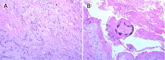
Fig. 2A. Histologic findings from the resected specimen (H&E, × 200). Invasive intermediate trophoblasts penetrated the myometrium through the superficial and deep portion. Fig. 2B. (H&E, × 200). In the perforation site near serosal side, there was a floating well developed villus admixed with fibrin material.
Figure & Data
References
Citations
Citations to this article as recorded by 
- Placenta accreta spectrum with severe morbidity: fibrosis associated with cervical-trigonal invasion
José M. Palacios-Jaraquemada, Álbaro José Nieto-Calvache, Rozi Aditya Aryananda, Nicolás Basanta, Clara Ivette Campos, Grace Ariani
The Journal of Maternal-Fetal & Neonatal Medicine.2023;[Epub] CrossRef
 PubReader
 ePub Link
 Cite
Cite

