 KOSIN UNIVERSITY COLLEGE OF MEDICINE
Articles
- Page Path
- HOME > Kosin Med J > Volume 30(1); 2015 > Article
-
Case Report
Tricuspid and pulmonary valve endocarditis associated with double-chambered right ventricle - Jin Cheol Myeong1, Jung Yeon Chin1, Jin Ho Choi2, Young Min Rah1, Jun Hyung Park1
-
Kosin Medical Journal 2015;30(1):81-85.
DOI: https://doi.org/10.7180/kmj.2015.30.1.81
Published online: January 20, 2015
1Department of Cardiology, Eulji University Hospital, Eulji University School of Medicine, Daejeon, Korea
2Department of Cardiovascular surgery, Eulji University Hospital, Eulji University School of Medicine, Daejeon, Korea
- Corresponding Author:Jin Cheol Myeong, Division of Cardiology, Eulji University Hospital, 95, Dunsan-Seoro, Seo-gu, Daejeon 302-120, Korea TEL: +82-10-9976-9992 FAX: +82-42-611-3183 Email: spyman2000@naver.com
• Received: September 18, 2014 • Accepted: October 15, 2014
Copyright © 2015 Kosin University School of Medicine Proceedings
This is an Open Access article distributed under the terms of the Creative Commons Attribution Non-Commercial License (http://creativecommons.org/licenses/by-nc/3.0) which permits unrestricted non-commercial use, distribution, and reproduction in any medium, provided the original work is properly cited.
- 880 Views
- 2 Download
Abstract
- We report a rare case of tricuspid valve and pulmonary valve endocarditis associated with a double-chambered right ventricle in an adult female with pulmonary artery aneurysm and septic pulmonary embolism by Streptococcus mitis. She was treated with aggressive antibiotic therapy followed by debridement of the infective lesion of tricuspid valve, pulmonary valve replacement using xenograft and resection of obstructing muscular bundles in right ventricle.
Figure 1.Initial electrocardiogram showing right ventricular hypertrophy with strain pattern, right axis deviation, and right atrial enlargement.

Figure 2.Pulmonary angio CT showing mild cardiomegaly with right heart enlargement and multiple distal pulmonary infarction (white arrow heads) with multiple pulmonary artery aneurysm (white arrows).
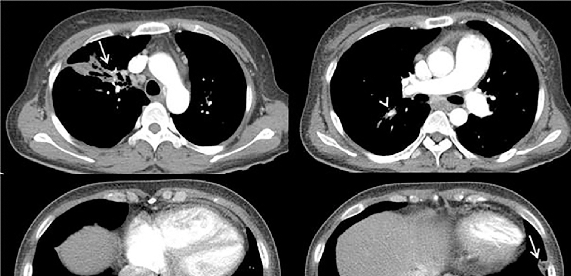
Figure 3.Transthoracic echocardiogram showing (A) continuous-wave Doppler tracing along the right ventricular outflow tract with peak gradient of 87 mmHg across the subinfundibulum (B) hypertrophied muscular bands dividing the right ventricle (white arrow) with D-shaped interventricular septum and large vegetation on pulmonary valve. LV: left ventricle, PV: pulmonary valve, RV: right ventricle.
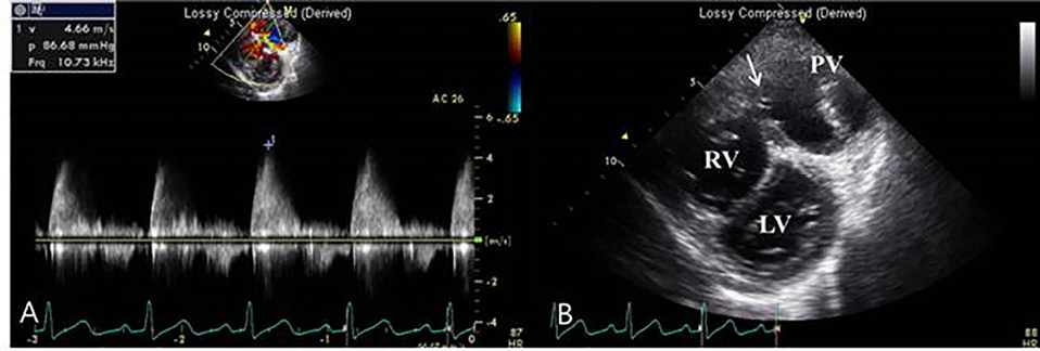
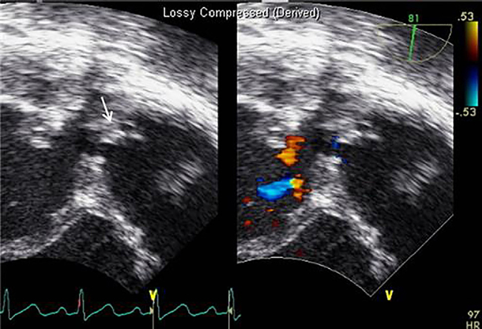
Figure 4.Transesophageal echocardiogram showing echogenic shaggy materials (white arrow) on hypertrophied muscular bands, suspicious vegetations.
- 1. Hoffman P, Wójcik AW, Rózan ′ski J, Siudalska H, Jakubowska E, Włodarska EK, et al. The role of echocardiography in diagnosing double chambered right ventricle in adults. Heart 2004;90:789–93.ArticlePubMedPMC
- 2. López-Pardo F, Aguilera A, Villa M, Granado C, Campos A, Cisneros JM. Double-chambered right ventricle associated with mural and pulmonic valve endocarditis: description of a clinical case and review of the literature. Echocardiography 2004;21:171–3.ArticlePubMed
- 3. Telagh R, Alexi-Meskishvili V, Hetzer R, Lange PE, Berger F, Abdul-Khaliq H. Initial clinical manifestations and mid- and longterm results after surgical repair of double-chambered right ventricle in children and adults. Cardiol Young. 2008;18:268–74.ArticlePubMed
- 4. Habib G, Hoen B, Tornos P, Thuny F, Prendergast B, Vilacosta I, et al. ESC Committee for Practice Guidelines.: Guidelines on the prevention, diagnosis, and treatment of infective endocarditis (new version 2009): the Task Force on the Prevention, Diagnosis, and Treatment of Infective Endocarditis of the European Society of Cardiology (ESC). Endorsed by the European Society of Clinical Microbiology and Infectious Diseases (ESCMID) and the International Society of Chemotherapy (ISC) for Infection and Cancer. Eur. Heart J 2009;30:2369–413.
- 5. Choi YJ, Park SW. Characteristics of double-chambered right ventricle in adult patients. Korean J Intern Med. 2010;25:147–53.ArticlePubMedPMC
- 6. Simpson Jr WF, Sade RM, Crawford FA, Taylor AB, Fyfe DA. Double chambered right ventricle. Ann Thorac Surg 1987;44:7–10.ArticlePubMed
- 7. Chaurasia AS, Nawale JM, Yemul MA. Double-Chambered Right Ventricle with Pulmonary Valve Endocarditis. Echocardiography 2013;30:167–70.Article
References
Figure & Data
References
Citations
Citations to this article as recorded by 
 PubReader
 ePub Link
 Cite
Cite


