KOSIN UNIVERSITY COLLEGE OF MEDICINE
Articles
- Page Path
- HOME > Kosin Med J > Volume 28(2); 2013 > Article
-
Case Report
A Tubercus Sclerosis Case Accompanied by Cystic Angiomyolipoma and Chronic Kidney Disease Diagnosed during Treatment for Acute Cerebral Infarction - Heejun Kim1, Inho Moh1, Da-Hye Jung1, Young-Ki Lee1, Ji-Young Woo2, Yul Lee2, Jung-Woo Noh1
-
Kosin Medical Journal 2013;28(2):177-182.
DOI: https://doi.org/10.7180/kmj.2013.28.2.177
Published online: January 19, 2013
1Department of Internal Medicine, Hallym Kidney Research Institute, Seoul, Korea
2Radiology, Hallym University College of Medicine, Seoul, Korea
- Corresponding author: Jung Woo Noh, Department of Internal Medicine, Kangnam Sacred Heart Hospital, Hallym University College of Medicine, 948-1 Daerim 1-dong, Yeoungdeungpo-gu, Seoul 150-950, Korea TEL: +82-2-829-5510 FAX: +82-2-848-9821 E-mail: jwn8671@unitel.co.kr
• Received: September 7, 2012 • Accepted: October 22, 2012
Copyright © 2013 Kosin University School of Medicine Proceedings
This is an Open Access article distributed under the terms of the Creative Commons Attribution Non-Commercial License (http://creativecommons.org/licenses/by-nc/3.0/) which permits unrestricted non-commercial use, distribution, and reproduction in any medium, provided the original work is properly cited.
- 1,081 Views
- 1 Download
Abstract
- Symptoms of tuberous scelrosis (TS) are mainly related with brain and kidneys. Seizure, mental retardation, other behavioral problems are dominant. A spectrum of renal tumors from benign angiomyolipoma (AML) to polycystic kidney disease, and rarely malignant renal cell carcinoma have been observed. Cystic AML is a rare phenotype of AML. No case of TS with renal cystic AML has been reported in Korea yet. And chronic kidney disease (CKD) in TS has been seldom reported. We experienced a TS case accompanied by renal cystic AML and CKD diagnosed in a 48-year-old female patient who was hospitalized for left side weakness and seizure under the diagnosis of acute cerebral infarction.
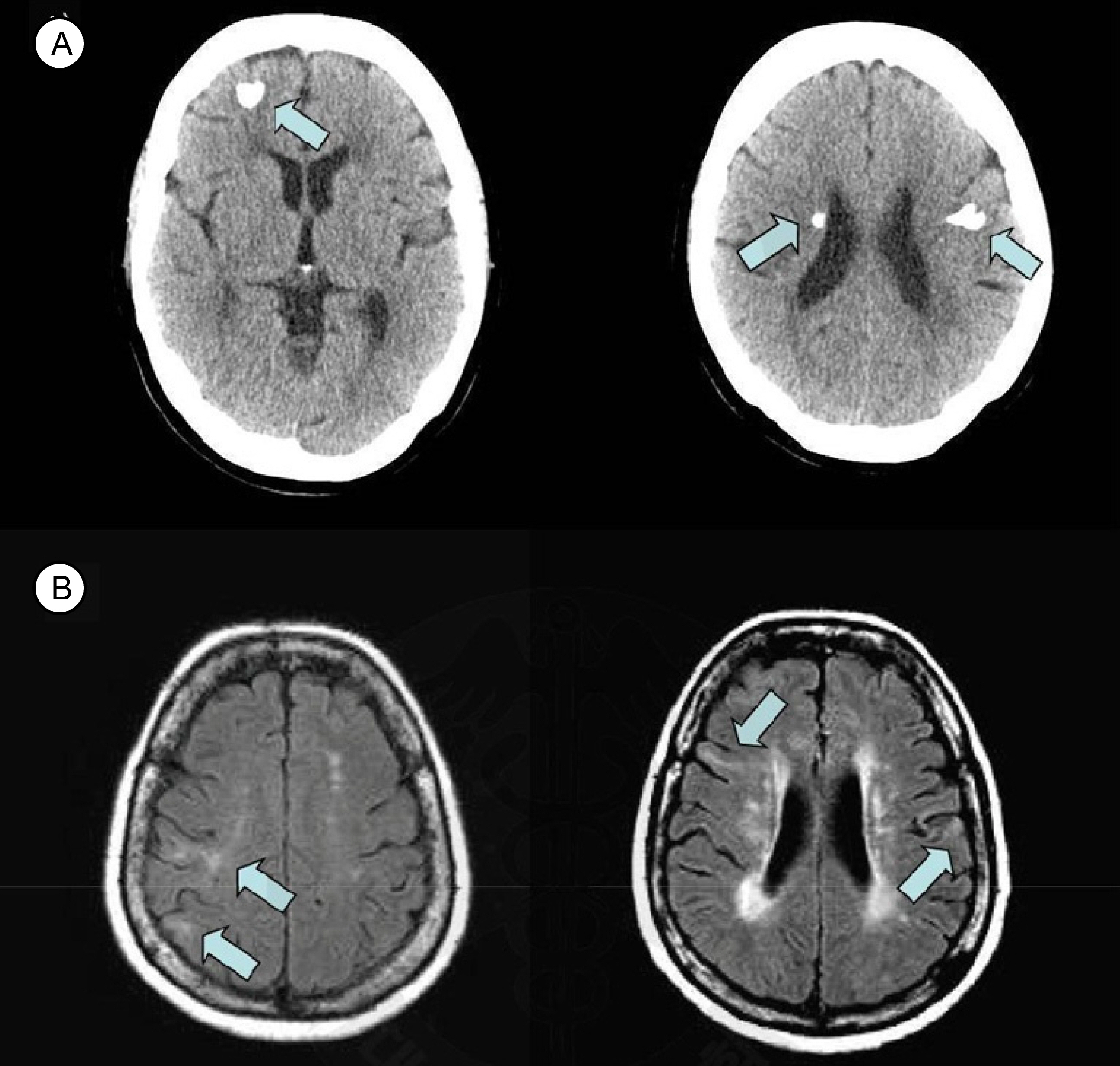
Fig. 2.(A) Brain CT shows multiple calcifications suggesting calcified tubers in right frontal periventricular area and left frontal subcortical white matter (arrows), (B) FLAIR image of MRI shows multifocal linear and wedge shaped high signal lesions suggesting cortical and subcortical tubers in both frontal cortex and subcortical white matter (arrows).

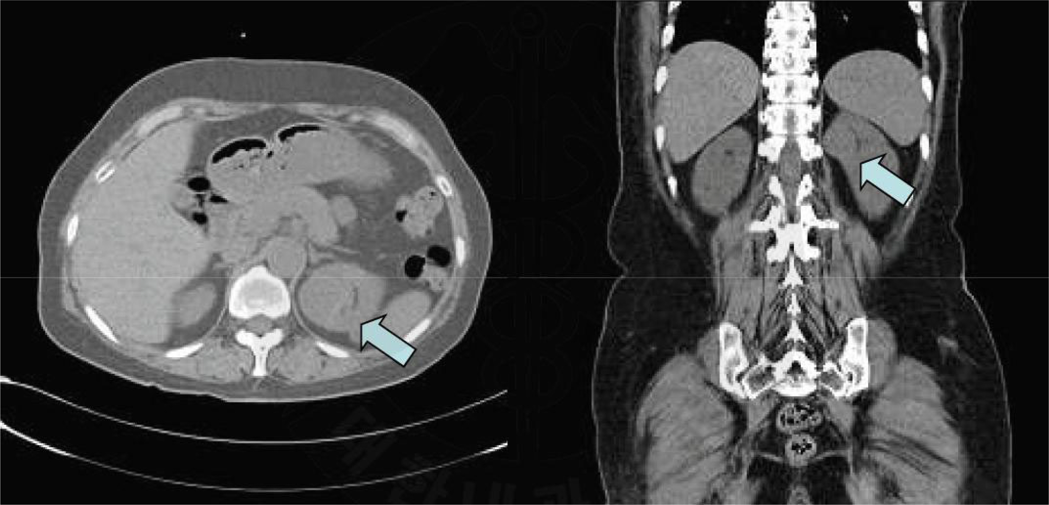
Fig. 4.(A) T2, (B) T1 fat setting, (C) T1 GRE, (D) T1. About 3.8 cm sized cystic mass with mixed components suggesting cystic angiomyolipoma and multiple cysts are seen (arrows) in MRI examination.

- 1.Narayanan V. Tuberous sclerosis complex: genetics to pathogenesis. Pediatr Neurol 2003;29:404–9.ArticlePubMed
- 2.Shepherd CW, Gomez MR, Lie JT, Crowson CS. Causes of death in patients with tuberous sclerosis. Mayo Clin Proc 1991;66:792–6.ArticlePubMed
- 3.Crino PB, Nathanson KL, Henske EP. The tuberous sclerosis complex. N Engl J Med 2006;355:1345–56.ArticlePubMed
- 4.Davis CJ, Barton JH, Sesterhenn IA. Cystic angiomyolipoma of the kidney: a clinicopathologic description of 11 cases. Mod Pathol 2006;19:669–74.ArticlePubMed
- 5.Yeo DM, Chung DJ, Kim TJ, Lee IK, Hahn ST, Lee JM. CT and MRI Findings of Angiomyolipoma with Epithelial Cysts of the Kidney: A Case Report. J Korean Soc Radiol 2010;63:173–6.Article
- 6.Neumann HP, Brüggen V, Berger DP, Herbst E, Blum U, Morgenroth A, et al. Tuberous sclerosis complex with end-stage renal failure. Nephrol Dial Transplant 1995;10:349–53.PubMed
- 7.Moon SH, Choi HJ, Yun UD, Yang DK, Woo YS, Chang KY, et al. Two Cases of Tuberous Sclerosis Patients with Renal Anomaly. Korean J Nephrol 2001;20:137–42.
- 8.Nelson CP, Sanda MG. Contemporary diagnosis and management of renal angiomyolipoma. J Urol 2002;168:1315–25.ArticlePubMed
- 9.Wiederholt WC, Gomez MR, Kurland LT. Incidence and Prevalence of tuberous sclerosis in Rochester, Minesota 1950-1982. Neurology 1985;35:600–3.ArticlePubMed
- 10.Hunt A. Lindenhaum RH. Tuberous sclerosis: A new estimate of prevalence within the Oxford region. J Med Genet 1984;21:272–7.PubMedPMC
- 11.Peter B. Crino, Katherine L. Nathanson, Elizabeth Petri Henske. The Tuberous Sclerosis Complex. N Engl J Med 2006;355:1345–56.ArticlePubMed
- 12.Larrode Pellicer P, Millán LF, Morales Asín F, Ayuso Blanco T, Bello Dronda S. Cerebral infarction and convulsive crises in a forme fruste of tuberous sclerosis. Rev Clin Esp 1986;179:477–8.PubMed
- 13.Kennelly MJ, Grossman HB, Cho KJ. Outcome analysis of 42 cases of renal angiomyolipoma. J Urol 1994;152:1988–91.ArticlePubMed
- 14.Jimenez RE, Eble JN, Reuter VE, Epstein JI, Folpe AL, de Peralta-Venturina M, et al. Concurrent angiomyolipoma and renal cell neoplasia: a study of 36 cases. Mod Pathol 2001;14:157–63.ArticlePubMed
References
Figure & Data
References
Citations
Citations to this article as recorded by 
 PubReader
 ePub Link
 Cite
Cite