KOSIN UNIVERSITY COLLEGE OF MEDICINE
Articles
- Page Path
- HOME > Kosin Med J > Volume 27(2); 2012 > Article
-
Case Report
A Case of Infective Endocarditis Occurred during Treatment for Infectious Spondylitis Accompanied by Peptostreptococcus Anaerobius Bacteremia - Byung Hee Lee, Myung Hee Lee, Sook Kyung Oh, Ji Young Seo, Joon Hoon Jeong, Jae Woo Lee
-
Kosin Medical Journal 2012;27(2):185-190.
DOI: https://doi.org/10.7180/kmj.2012.27.2.185
Published online: December 27, 2012
1Department of Internal Medicine, Wallace Memorial Baptist Hospital, Busan, Korea.
2Department of Cardiology, College of Medicine, Kosin University, Busan, Korea.
- Corresponding Author: Joon Hoon Jeong, Department of Internal Medicine, Wallace Memorial Baptist Hospital, 374-75 Namsan-dong, Geumjeong-gu, Busan 609-728, Korea. TEL: +82-51-580-1202, FAX: +82-51-583-7114, jjhoon69@yahoo.co.kr
• Received: July 10, 2012 • Revised: September 14, 2012 • Accepted: October 17, 2012
Copyright © 2012 Kosin University College of Medicine
- 986 Views
- 3 Download
- 1 Crossref
Abstract
- It is necessary to distinguish between pyogenic and tuberculous spondylitis of infectious spondylitis, if it is pyogenic spondylitis, antimicrobial therapy should be directed against an identified microorganism and clinical assessment should be done at 4 weeks. But if microorganism is a anaerobic bacteria, especially Peptostreptococcus anaerobius, combination antibiotic therapy should be considered bacause it may be a component of mixed infections as a passenger and have abilities to induce abscesses, other bacterial growth as a synergy effect. In addition, echocardiography may be necessary because pyogenic spondylitis is associated with infective endocarditis about 12%. We report a 64-year-old man who was treated for infectious spondylitis accompanied by Peptostreptococcus anaerobius bacteremia, but had to undergo heart surgery because an attack of infective endocarditis with systemic embolism during hospitalization.
- 1. Kim YI, Kim SE, Jang HC, Jung SI, Song SK, Park KH. Analysis of the Clinical Characteristics and Prognostic Factors of Infectious Spondylitis. Infect Chemother 2011;43:48–54.Article
- 2. Koo KH, Lee HJ, Chang BS, Yeom JS, Park KW, Lee CK. Differential Diagnosis between Tuberculous Spondylitis and Pyogenic Spondylitis. J Korean Soc Spine Surg 2009;16:112–121.Article
- 3. Gouliouris T, Aliyu SH, Brown NM. Spondylodiscitis: update on diagnosis and management. J Antimicrob Chemother 2010;65:iii11–iii24.ArticlePubMed
- 4. Murdoch DA. Gram-positive anaerobic cocci. Clin Microbiol Rev 1998;11:81–120.ArticlePubMedPMC
- 5. Zimmerli W. Clinical practice. Vertebral osteomyelitis. N Engl J Med 2010;362:1022–1029.ArticlePubMed
- 6. Cottle L, Riordan T. Infectious spondylodiscitis. J Infect 2008;56:401–412.ArticlePubMed
- 7. Minces LR, Shields RK, Sheridan K, Ho KS, Silveira FP. Peptostreptococcus infective endocarditis and bacteremia. Analysis of cases at tertiary medical center and review of the literature. Anaerobe 2010;16:327–330.ArticlePubMed
- 8. Könönen E, Bryk A, Niemi P, Kanervo-Nordström A. Antimicrobial susceptibilities of peptostreptococcus anaerobius and the newly described peptostreptococcus stomatis isolated from various human sources. Antimicrob Agents Chemother 2007;51:2205–2207.ArticlePubMedPMC
- 9. Lee KY, Chong YS, Jeong SH, Xu XS, Kwon OH. Emerging Resistance of Anaerobic Bacteria to Antimicrobial Agents in South Korea. Clin Infect Dis 1996;23:S73–S77.ArticlePubMed
- 10. The Korean Society of Infectious Diseases. Korean Society for Chemotherapy. The Korean Society of Clinical Microbiology. The Korean Society of Cardiology. The Korean Society for Thoracic and Cardiovascular Surgery. Clinical Guideline for the Diagnosis and Treatment of Cardiovascular Infection. Infect Chemother 2011;43:129–177.Article
References
Fig. 1Spine MRI shows diffuse contrast enhancement of prevertebral soft tissue and endplate at L5-S1 (arrow).
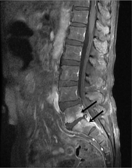
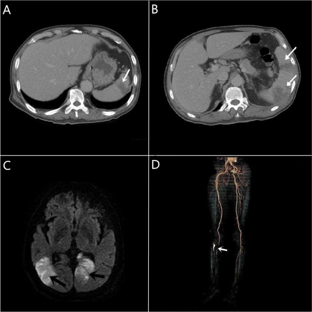
Fig. 2Septic emboli identified according to imaging modalities (arrow). (A) Contrast-enhanced abdominal CT shows wedge shaped low attenuated lesion at spleen. (B) Aggravated state of spleen after 8 weeks. (C) Brain MRI shows acute ischemic infarct in both occipital lobe. (D) CT angiography of femoral artery shows focal segmental occlusion at distal portion of right popliteal artery.

Figure & Data
References
Citations
Citations to this article as recorded by 
- Endocarditis due to Gram Positive Anaerobic Cocci: First report of Peptoniphilus indolicus endocarditis and literature review
Julie Lourtet-Hascoet, Sébastien Hascoet, Jean-Louis Galinier, Benoît Fontenel, Benoît Monteil, Eric Bonnet
Clinical Infection in Practice.2021; 11: 100073. CrossRef
 PubReader
 ePub Link
 Cite
Cite