Successful Removal of 15-year-old Pacemaker Leads by Weight and pulley method
Article information
Abstract
Extraction of old pacemaker leads remains a complex procedure owing to fibrotic encapsulation and lead adhesions. We report a case of extraction of 15-year-old pacemaker leads by weight and pulley method. A 81-year-old man presented with exposed pacemaker leads out of body with purulent discharge from a pacemaker insertion site. He inserted DDD (dual chamber pacing, dual chamber sensing dual function) pacemaker implantation 15 years ago for SSS. Previously pacemaker battery was removed 3 years ago due to recurrent infection of pacemaker scar site. We extracted the pacemaker leads by weight and pulley method successfully without any complications.
The number of implanted cardiac implantable electronic devices has increased recent years.1 This trend is due to wider range of indication.23 Despite improved lead performance, an increasing number of device patients, better life expectancy, more leads per patient, new indications, new types of devices, as well as device and lead recalls strongly increased the need for lead extraction. Every year more than 10,000 – 15,000 patients are produced lead extraction in worldwide.4
The extraction of chronically implanted pacemaker leads is a challenging problem. Manual traction or methods such as weight and pulley or forceps-assisted traction were often found to be both unsuccessful and prone to complication.4
Recently, the development of new extraction techniques has renewed the interest in this particular field. But, the associated high costs and complications are still remained problem.
We describe a case of lead extraction using weight and pulley method.
CASE
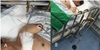
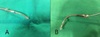
A 81-year-old man presented with exposed pacemaker leads out of his body with purulent discharge from a insertion site in the left pectoral area (Fig. 1). He inserted DDD (dual chamber pacing, dual chamber sensing dual function, Guidant model 1230, Boston scientific, USA) pacemaker implantation 15 years ago for sick sinus syndrome (SSS). Previously pacemaker battery was removed 3 years ago due to recurrent infection of pacemaker scar site. At that time, we planned to remove the remained pacemaker leads, but he did not visit hospital since then.
exposed pacemaker leads out of his body with purulent discharge from a insertion site in the left pectoral area
At admission, the patient's blood pressure was 110/70 mmHg and heart rate was 67 and chronic-ill looking appearance. Two pacemaker leads were well positioned in right atrium and right ventricle in initial chest x-ray (Fig. 2A).
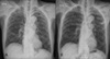
Chest x-ray at admisstion and post extracted lead. (A) initial chest x-ray. atrial and ventricular lead was placed correctly, but opposite leads were exosed out of body. (B) After atrial and ventricular leads was applied weight pully method, the leads were extracted.
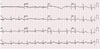
Electrocardiogram (EKG) recorded sinus rhythm with a heart rate 70 bpm (Fig. 3). The transthoracic echocardiography (TTE) showed that the ejection fraction was 67% and no definite vegetation or thrombus were seen.
Laboratory analysis revealed a white blood cell count of 16,470/L, a neutrophil count of 91.0%, Hgb 12.9g/dL, platelet 178,000/L, Procalcitonin 3.47ng/mL, C-reactive protein levels of 15.74mg/dL and initial blood culture growing Methicillin sensitive staphylococcus areus.
Because Laboratory test results were septic condition due to pacemaker lead infection, we decided to remove pacemaker leads by surgical operation. But the patient refused surgical operation, we planned to remove pacemaker leads by weight and pulley method.
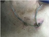
The force applied during ventricle lead extraction was 1 Ib. After 1 day, ventricle lead was totally extracted (Fig. 4).
Weight pully method. (A) ventricular lead tip was extracted by forcep (B) The force applied during ventricular lead extraction was 1 Ib. The person on strict bed rest must remain in bed at all times.
The force applied during atrium lead extraction was 1 Ib, but the lead was not extracted for 2 days. After traction force increase from 1 Ib to 2 Ib, prolonged and graded traction has been introduced. Two days later, the atrium lead was totally extracted (Fig. 5A, B).
The extreaced leads. (A) ventricular lead (B) atrial lead
Follow-up TTE showed that the ejection fraction was 60% and no complication. Vital sign was stable (blood pressure 100/60 mmHg, heart rate 68) and laboratory analysis revealed a white blood cell count of 7,080/µL, a neutrophil count of 70.2%, Hgb 13.1g/dL, platelet 257,000/L, C-reactive protein levels of 1.69 mg/dL, negative wound culture and 5-day negative follow-up blood culture. Two pacemaker leads were totally extracted in chest x-ray (Fig. 2B).
Five days after discharge, the patient's blood pressure was 110/70 mmHg and heart rate was 54, EKG showed a sinus rhythm with no ST-segment change.
DISCUSSION
There are many ways to remove the pacemaker lead.
1. manual traction without tools
2. traction medicated by some sort of weight or by application of a clamp to the stretched lead
3. mechanical sheaths, with or without the use of a locking stylet
4. laser-assisted lead extraction, with or without the use of a locking stylet
5. open chest extraction, with or without transvenous extraction tools
6. transthoracic extraction using a paraternal, subxyphoid or intercostals approach.5
Although newly developed techniques such as electrosurgical or laser sheath are in clinical use, they have not been introduced in Korea, and the conventional technique using locking stylet and dilator sheath is still useful if performed by experienced operator.6 Compare to conventional technique, manual extraction dose not usually used because of complications (e.g. ventricle avulsion or rupture).
Pacemaker lead extraction is generally considered to be a difficult and high-risk procedures by manual traction using weight pulley method, but there are also complications in using locking stylets and sheaths because of direct injury to adhesion site of major vein. And systemic lead infection seems to counteract or dissolve fibrotic adherences.
Although there is interoperator variability in the forces exerted, are typically around 11N and considered safe in manual traction procedures. The currently applied traction forces are in the same range as those used during the continuous traction era when weights of up to 3 Ibs(~12N) over a maximum of 7 days were recommended.7 The force applied in this case is 1~2 Ib and it was effective.
With increasing rates of pacemaker implantation, pacemaker infection rates have risen in parallel.8 In case of infection, pacemaker removal and lead extraction are necessary.
Although Weight and pulley method is difficult and high-risk procedures, this case shows Weight and pulley method can be alternative lead extraction method of the cardiac implantable electronic devices when patients refuse procedure or surgical operation associated with high cost. Because of this reason, we report this case.