Biliary Cystadenoma Causing Esophageal Varices
Article information
Abstract
Abstract
Biliary cystadenomas are benign but potentially malignant cystic neoplasm. The preferred treatment is radical resection because it is difficult to differentiate a benign from a malignant biliary cystadenoma. A 40 year-old woman presented with moderate abdominal discomfort. Esophageal varix was found up to mid-esophagus on endoscopy. She has no prior history of liver disease or chronic alcohol ingestion. About 15cm sized biliary cystadenoma was diagnosed by ultrasonography, computed tomography and magnetic resonance imaging. Serum level of bilirubin, alanine aminotransferase, alkaline phosphatase, gamma-glutamyl transpeptidase and tumor marker were elevated. The patient underwent US-guided aspiration. Tumor markers from the aspirated fluid are increased. Left hepatectomy was performed to completely remove the cyst. Histology of the resected specimen confirmed a biliary cystadenoma of the liver with ovary-like stroma. Without prior history of liver disease or chronic alcoholic ingestion, incidental finding of esophageal varix could show an important clue for diagnosis of biliary cystadenoma.

Abdomen ultrasonography shows a large cystic mass with septae in liver left lobe.
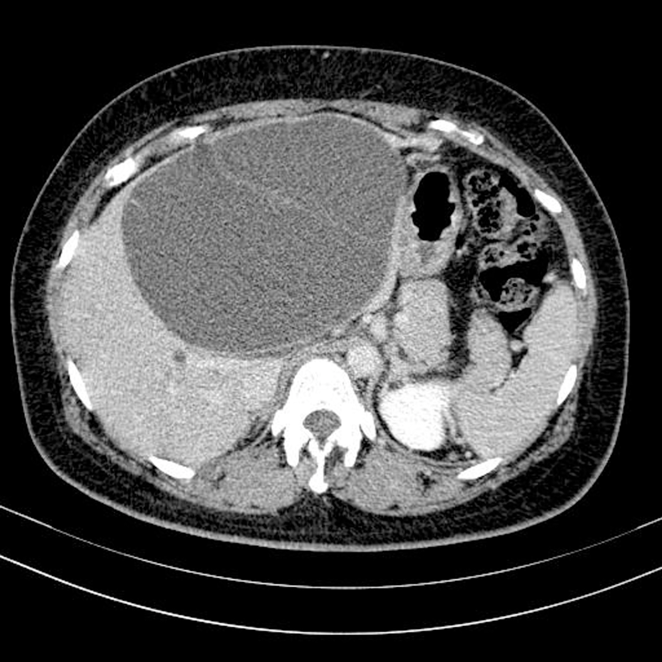
Computed tomography shows a large cystic lesion with multiple-enhancing septae (arrow).
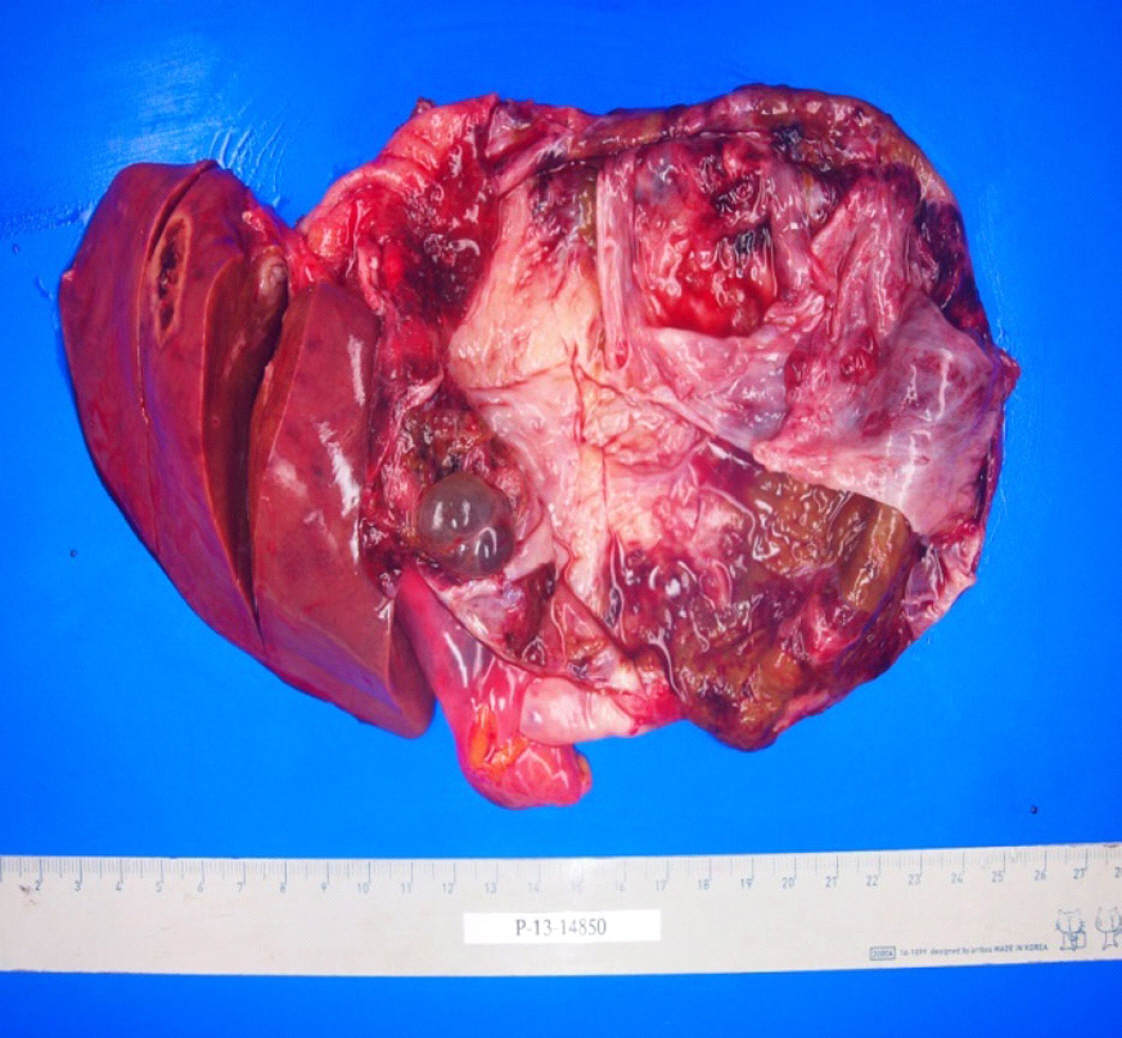
Postoperative picture shows large thick walled cystic lesion in left lobe of liver.
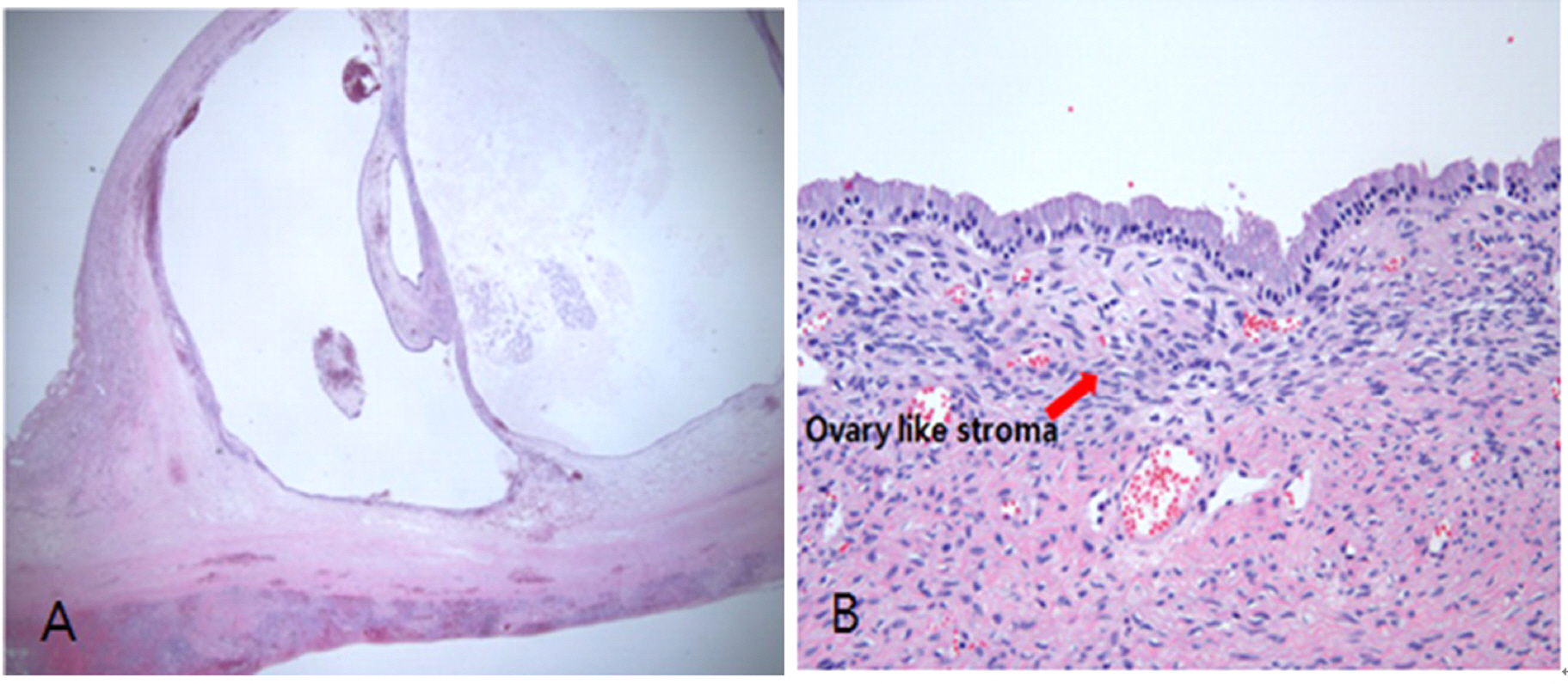
Cyst histology typical of a mucinous cystadenoma(A) Biliary cyst shows ovary like stroma (H &E stain, x400). (B) Arrow demonstrating ovary like stroma.

Endoscopy image before operation demonstrating esophageal varix upto midesophagus.(A) Follow up endoscopy after lobectomy shows disappearance of esophageal varix.