 KOSIN UNIVERSITY COLLEGE OF MEDICINE
Articles
- Page Path
- HOME > Kosin Med J > Volume 27(1); 2012 > Article
-
Case Report
A Case of Henoch Schönlein Purpura with Gastrointestinal Bleeding Due to Jejunal Ulcer by Capsule Endoscopy - Yong Kang Lee, Tak Keun Oh, Ara Choi, Ji Hoon Lee, Mi Na Kim, Sung Pil Hong
-
Kosin Medical Journal 2012;27(1):45-49.
DOI: https://doi.org/10.7180/kmj.2012.27.1.45
Published online: June 11, 2012
1Department of Internal Medicine, College of Medicine, Yonsei University, Seoul, Korea.
2Division of Gastroenterology, College of Medicine, Yonsei University, Seoul, Korea.
- Corresponding Author: Sung Pil Hong, Department of Internal Medicine, College of Medicine, Yonsei University, 250 Seongsanno, Seodaemun-gu, Seoul 120-752, Korea. TEL: 010-8940-5775, FAX: 02) 393-6884, sphong@yuhs.ac
• Received: September 28, 2011 • Revised: November 9, 2011 • Accepted: November 28, 2011
Copyright © 2012 Kosin University College of Medicine
- 1,132 Views
- 2 Download
- 1 Crossref
Abstract
-
- Henoch-Schönlein purpura (HSP) is the most common form of systemic vasculitis in children. Palpable purpura, arthralgia, arthritis, abdominal pain and renal involvement are the major clinical manifestations. Gastrointestinal involvement is related with abdominal pain and bleeding.
- We described a 71 year-old female experienced acute exacerbation of HSP presented with gastrointestinal bleeding. She was hospitalized for hematemesis and diagnosed duodenitis by esophagogastroduonenoscopy (EGD). Duodenitis was improved at EGD checked in 7 days. She still complained of melena and abdominal pain. There were no abnormal findings at sigmoidoscopy. Jejunal ulcer and purpura were diagnosed by capsule endoscopy. Symptoms were relieved after administration of systemic steroid. But she needed renal replacement therapy for 3 months.
- Small bowel ulcer diagnosed by capsule endoscopy in patients with HSP was rarely described in Korean literature. This case suggests that capsule endoscopy have a role in diagnosis of small bowel ulcer and its severity in HSP with gastrointestinal symptom.
- 1. Gardner-Medwin JM, Dolezalova P, Cummins C, Southwood TR. Incidence of Henoch-Schonlein purpura, Kawasaki disease, and rare vasculitides in children of different ethnic origins. Lancet 2002;360:1197–1202.ArticlePubMed
- 2. Pillebout E, Thervet E, Hill G, Alberti C, Vanhille P, Nochy D. Henoch-Schonlein Purpura in adults: outcome and prognostic factors. J Am Soc Nephrol 2002;13:1271–1278.PubMed
- 3. Nishiyama R, Nakajima N, Ogihara A, Oota S, Kobayashi S, Yokoyama K, et al. Endoscope images of Schonlein-Henoch purpura. Digestion 2008;77:236–241.ArticlePubMedPDF
- 4. Stancanelli B, Vita A, Vinci M, Magnano A, Purrello F. Bleeding of small bowel in Henoch-Schonlein syndrome: the successful diagnostic role of video capsule endoscopy. Am J Med 2006;119:82–84.ArticlePubMed
- 5. Lee GW, Cheon YK, Kim HJ, Lee SH, Cho JY, Shim CS. A Case of Henoch-Schonlein Purpura with Small Bowel Hemorrhage Diagnosed by Capsule Endoscopy. Korean J Gastrointest Endosc 2004;28:317–320.
- 6. Choi WH, Kim NH, Jung ES, Yoon SG, Park JS, Bae WK, et al. A case of terminal ileal ulcer of Henoch-Schonlein purpura treated with high dose steroid. Korean J Gastroenterol 2007;50:324–327.PubMed
- 7. Chang WL, Yang YH, Lin YT, Chiang BL. Gastrointestinal manifestations in Henoch-Schonlein purpura: a review of 261 patients. Acta Paediatr 2004;93:1427–1431.ArticlePubMed
- 8. Liu K, Kaffes AJ. Review article: the diagnosis and investigation of obscure gastrointestinal bleeding. Aliment Pharmacol Ther 2011;34:416–423.ArticlePubMed
- 9. Ichikawa R, Hosoe N, Imaeda H, Takabayashi K, Bessho R, Ida Y, et al. Evaluation of small-intestinal abnormalities in adult patients with Henoch-Schonlein purpura using video capsule. Endoscopy 2011;43:E162–E163.ArticlePubMed
- 10. Tobino K, Shimizu Y, Miura S, Sugawara K, Takeda K, Tomino Y. Severe erosive lesions in the digestive tract of patients with Henoch-Schönlein Purpura (HSP) and its impact on prognosis - presentation of two cases and statistical review of adult-onset Japanese HSP. Clin Nephrol 2011;75:Suppl 1. 47–55.PubMed
References
Fig. 1Esophagogastroduodenoscopy (EGD) findings. (A) EGD showed mild chronic superficial gastritis (B) EGD showed severe duodenitis from bulb to 2nd portion of duodenum.
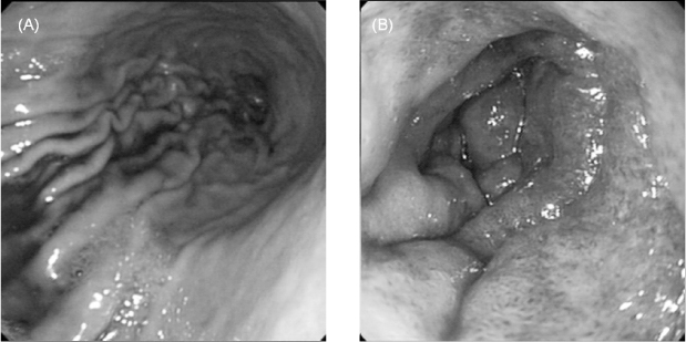
Fig. 2Capsule endoscopy showed multiple ulcer and erosions, multiple erythematous lesion and ulcer at Jejunum
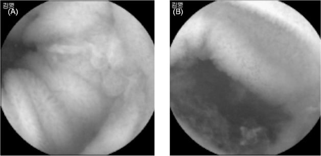
Figure & Data
References
Citations
Citations to this article as recorded by 
- A practical approach for small bowel bleeding
Sung Eun Kim, Hyun Jin Kim, Myeongseok Koh, Min Cheol Kim, Joon Sung Kim, Ji Hyung Nam, Young Kwan Cho, A Reum Choe
Clinical Endoscopy.2023; 56(3): 283. CrossRef
 PubReader
 ePub Link
 Cite
Cite


