 KOSIN UNIVERSITY COLLEGE OF MEDICINE
Articles
- Page Path
- HOME > Kosin Med J > Volume 30(2); 2015 > Article
-
Original Article
Mucinous precursor lesions of mucinous carcinoma in breast: Incidence and histopathologic features - Min Jung Jung, Young Ok Kim
-
Kosin Medical Journal 2015;30(2):131-139.
DOI: https://doi.org/10.7180/kmj.2015.30.2.131
Published online: January 20, 2015
Department of Pathology, College of Medicine, Kosin University, Busan, Korea
- Corresponding Author: Young Ok Kim, Department of Pathology, College of Medicine, Kosin University, 262, Gamcheon-ro, Seo-gu, Busan 49267, Korea Tel: 82-51-990-6325 Fax: 82-51-990-3080 E-mail: suajoon@ns.kosinmed.or.kr
Copyright © 2015 Kosin University School of Medicine Proceedings
This is an Open Access article distributed under the terms of the Creative Commons Attribution Non-Commercial License (http://creativecommons.org/licenses/by-nc/3.0) which permits unrestricted non-commercial use, distribution, and reproduction in any medium, provided the original work is properly cited.
- 851 Views
- 1 Download
Abstract
-
Objectives
- Columnar cell lesion (CCL), atypical ductal hyperplasia (ADH) and ductal carcinoma in situ (DCIS) may be premalignant lesion of mammary invasive carcinoma. A few recent investigators reported that the precursor lesions exhibited mucin production and they might be potential precursor lesion for mucinous carcinoma (mCA). This study aims to investigate the incidence and histopathologic characteristics of mucinous precursor lesions, including mucinous DCIS (mDCIS) and mucinous CCL (mCCL).
-
Methods
- We retrospectively analyzed invasive carcinomas with mucin. Cases were grouped into three subgroups: pure mCA, mixed mCA, and invasive carcinoma of no special type with mucin production (IC of NST-m). Precursor lesions were evaluated with PAS and alcian blue staining.
-
Results
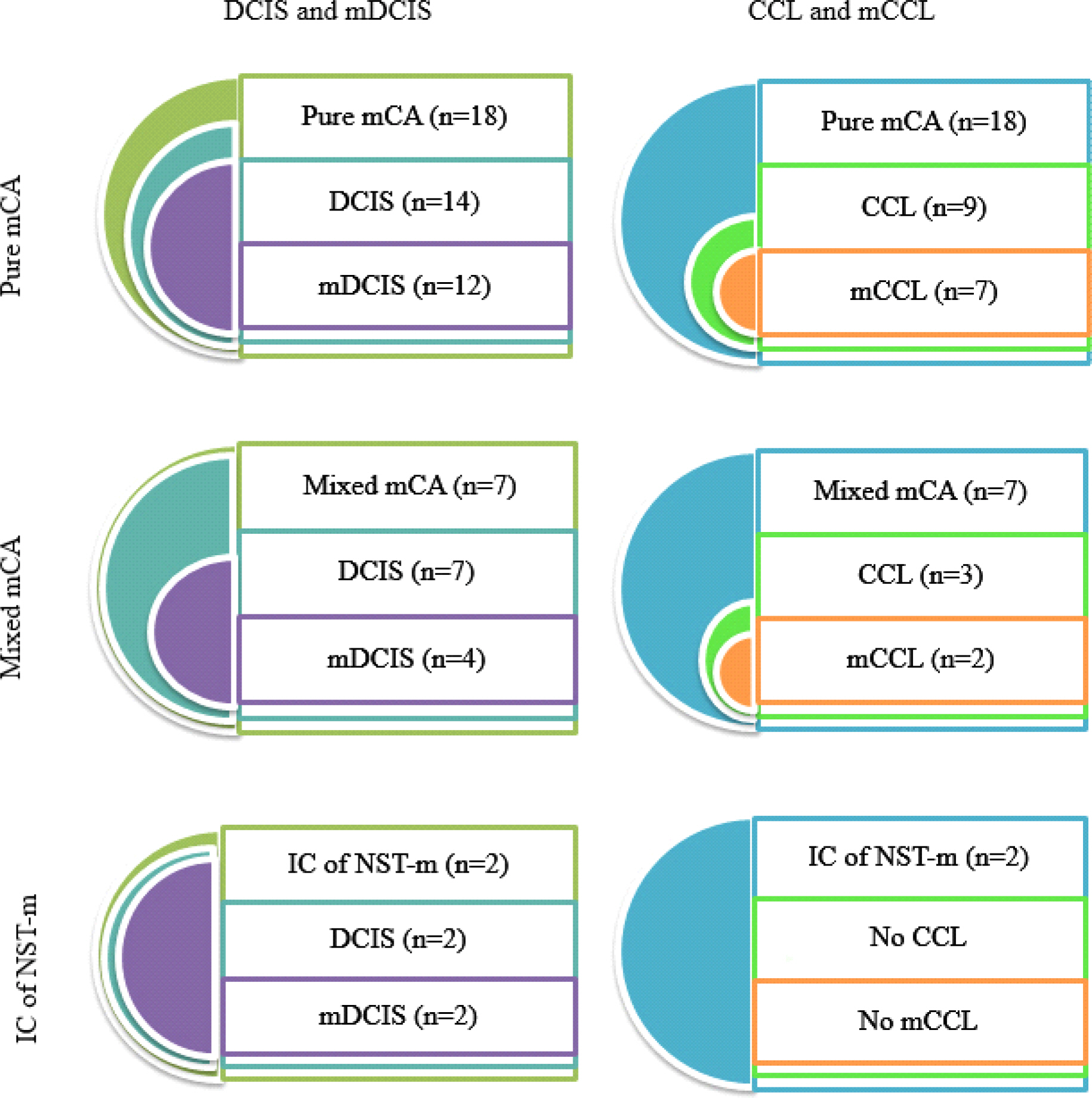
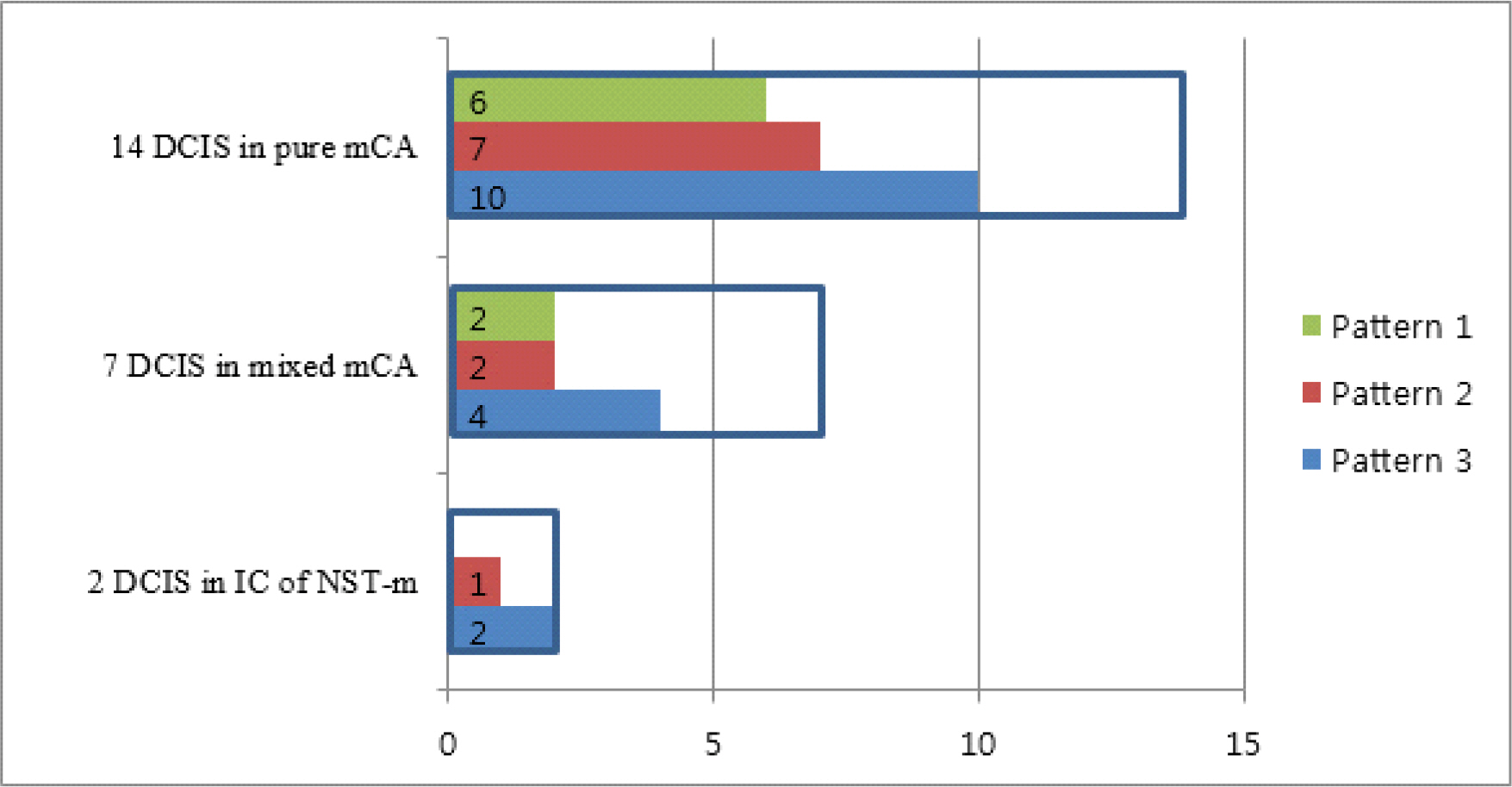
- Total 27 cases of invasive carcinoma with mucin were analysed and classified as 18 pure mCA, 7 mixed mCA, and 2 IC of NST-m. mDCISs were found in 12 pure mCA, 4 mixed mCA and 2 IC of NST-m. mCCLs were found in 7 pure mCA and 2 mixed mCA. Majority of mucin was identified in both cytoplasm and ductal lumen, while some tumors exhibited only cytoplasmic mucin. We also observed three patterns of mDCIS classifiable by location of mucin and architecture of tumor cells.
-
Conclusions
- Cytoplasmic mucin suggested that mucinous feature of precursor lesions in the vicinity of mCA might not be a passive morphologic finding but be involved in development of mCA.

mCA, mucinous carcinoma; IC of NST-m, invasive carcinoma of no special type with mucin production; Min, minimal; Max, maximal; M, mastectomy; E, excision; E+M, excision with subsequent mastectomy; +, present; -, absent; IC with SRC diff, invasive carcinoma with signet-ring cell differentiation; IC of NST, invasive carcinoma of no special type
mCA, mucinous carcinoma; IC of NST-m, invasive carcinoma of no special type with mucin production; DCIS, ductal carcinoma in situ; mDCIS, mucinous ductal carcinoma in situ; CCL, columnar cell lesion; mCCL, mucinous columnar cell lesion; +, present; -, absent; NA, not available; L, ductal lumen; C, cytoplasm
- 1. Abdel-Fatah TM, Powe DG, Hodi Z, Reis-Filho JS, Lee AH, Ellis IO. Morphologic and molecular evolutionary pathways of low nuclear grade invasive breast cancers and their putative precursor lesions: further evidence to support the concept of low nuclear grade breast neoplasia family. Am J Surg Pathol 2008;32:513–23.ArticlePubMed
- 2. Simpson PT, Gale T, Reis-Filho JS, Jones C, Parry S, Sloane JP, et al. Columnar cell lesions of the breast: the missing link in breast cancer progression? A morphological and molecular analysis. Am J Surg Pathol 2005;29:734–46.PubMed
- 3. Sunil R. Lakhani IOE, Stuart J Schinitt, Puay Hoon Tan, Marc J. van de Vijver. WHO classification of tumours of the breast. 4th Edition ed. Fred T. Bosman ESJ, Sunil R. Lakhani, Hiroko Ohgaki, editor. Geneva: International Agency for Research on Cancer; 2012.
- 4. Go EM, Tsang JY, Ni YB, Yu AM, Mendoza P, Chan SK, et al. Relationship between columnar cell changes and low-grade carcinoma in situ of the breast–a cytogenetic study. Hum Pathol 2012;43:1924–31.ArticlePubMed
- 5. Moinfar F, Man YG, Bratthauer GL, Ratschek M, Tavassoli FA. Genetic abnormalities in mammary ductal intraepithelial neoplasia-flat type ("clinging ductal carcinoma in situ"): a simulator of normal mammary epithelium. Cancer 2000;88:2072–81.ArticlePubMed
- 6. Schnitt SJ. The diagnosis and management of preinvasive breast disease: flat epithelial atypia–classification, pathologic features and clinical significance. Breast Cancer Res 2003;5:263–8.ArticlePubMedPMCPDF
- 7. Abdel-Fatah TM, Powe DG, Hodi Z, Lee AH, Reis-Filho JS, Ellis IO. High frequency of coexistence of columnar cell lesions, lobular neoplasia, and low grade ductal carcinoma in situ with invasive tubular carcinoma and invasive lobular carcinoma. Am J Surg Pathol 2007;31:417–26.ArticlePubMed
- 9. Gadre SA, Perkins GH, Sahin AA, Sneige N, Deavers MT, Middleton LP. Neovascularization in mucinous ductal carcinoma in situ suggests an alternative pathway for invasion. Histopathology 2008;53:545–53.ArticlePubMed
- 10. Kryvenko ON, Chitale DA, Yoon J, Arias-Stella J 3rd, Meier FA, Lee MW. Precursor lesions of mucinous carcinoma of the breast: analysis of 130 cases. Am J Surg Pathol 2013;37:1076–84.PubMed
- 11. O'Connell JT, Shao ZM, Drori E, Basbaum CB, Barsky SH. Altered mucin expression is a field change that accompanies mucinous (colloid) breast carcinoma histogenesis. Hum Pathol 1998;29:1517–23.ArticlePubMed
- 12. Di Saverio S, Gutierrez J, Avisar E. A retrospective review with long term follow up of 11,400 cases of pure mucinous breast carcinoma. Breast Cancer Res Treat 2008;111:541–7.ArticlePubMed
- 13. Matsukita S, Nomoto M, Kitajima S, Tanaka S, Goto M, Irimura T, et al. Expression of mucins (MUC1, MUC2, MUC5AC and MUC6) in mucinous carcinoma of the breast: comparison with invasive ductal carcinoma. Histopathology 2003;42:26–36.ArticlePubMed
- 14. Komaki K, Sakamoto G, Sugano H, Morimoto T, Monden Y. Mucinous carcinoma of the breast in Japan. A prognostic analysis based on morphologic features. Cancer 1988;61:989–96.ArticlePubMed
 PubReader
 ePub Link
 Cite
Cite


