 KOSIN UNIVERSITY COLLEGE OF MEDICINE
Articles
- Page Path
- HOME > Kosin Med J > Volume 28(2); 2013 > Article
-
Original Article
Application of Digital Infrared Thermographic Imaging (DITI) in the Monitoring of Change of Skin Temperature about Vascular Supply of Lower Abdominal Axial Flap in the Rabbit - Hyun nam Choi, Jin Hyung Park, Yea Sik Han, Sin Rak Kim, Han Kyeol Kim
-
Kosin Medical Journal 2013;28(2):131-136.
DOI: https://doi.org/10.7180/kmj.2013.28.2.131
Published online: January 19, 2013
Department of Plastic and Reconstructive Surgery, College of Medicine, Kosin University, Busan, Korea
- Corresponding author: Yea Sik Han, Department of Plastic Surgery, College of Medicine, Kosin University, 34 Amnam-dong, Seo-gu, Busan, 602-702, Korea TEL: +82-51-990-6131 FAX: +82-51-990-3005 E-mail: hanplastic1@naver.com
• Received: February 8, 2013 • Accepted: July 23, 2013
Copyright © 2013 Kosin University School of Medicine Proceedings
This is an Open Access article distributed under the terms of the Creative Commons Attribution Non-Commercial License (http://creativecommons.org/licenses/by-nc/3.0/) which permits unrestricted non-commercial use, distribution, and reproduction in any medium, provided the original work is properly cited.
- 918 Views
- 2 Download
Abstract
-
Methods
- Eight male New Zealand white rabbits with average weight of 3kg were used. A 10 x 10 cm unipedicled fasciocutaneous island flap was elevated based on the left superficial inferior epigastric vessel. The surface temperatures on designed flap were checked with DITI for 24 hours after the operation. On 14th day after the operation, the surviving area was measured and compared with DITI image which was taken on 24 hours after the operation using digital analysis software ImageJ. Statistical analysis was evaluated by paired T-test
-
Results
- On DITI image 24 hours after the flap elevation, distal portion of the flap showed remarkable color change. The average percentage and the standard deviation of the survival area of the flap which is predicted by DITI and the average percentage and the standard deviation of the survival area of the flap which was actually measured 2 weeks after flap elevation were 55.3 (16.6), 56.2 (18.0), respectively. This shows no significant difference between the two.
-
Objectives
- Monitoring viability of flap is important. The flap survival depends on the vascularity of the flap, on which the skin temperature depends. The authors applied digital infrared thermographic imaging (DITI) for monitoring the vascular supply of the flap and for the prediction of the prognosis of the flap survival.
-
Methods
- Eight male New Zealand white rabbits with average weight of 3kg were used. A 10 x 10 cm unipedicled fasciocutaneous island flap was elevated based on the left superficial inferior epigastric vessel. The surface temperatures on designed flap were checked with DITI for 24 hours after the operation. On 14th day after the operation, the surviving area was measured and compared with DITI image which was taken on 24 hours after the operation using digital analysis software ImageJ. Statistical analysis was evaluated by paired T-test
-
Results
- On DITI image 24 hours after the flap elevation, distal portion of the flap showed remarkable color change. The average percentage and the standard deviation of the survival area of the flap which is predicted by DITI and the average percentage and the standard deviation of the survival area of the flap which was actually measured 2 weeks after flap elevation were 55.3 (16.6), 56.2 (18.0), respectively. This shows no significant difference between the two.
-
Conclusions
- This study shows that DITI system could be used in evaluation of flap vascularity with ease, quickness and safety for patient and flap. Thus, it could be used clinically for the prediction of flap survival.
Fig. 1.Design and elevation of fasciocutaneous island flap. (A) Preoperative marking of the flap, (B) Intraoperative view of 10 x 10 cm sized left superficial inferior epigastric artery based fasciocutaneous island flap elevation.

Fig. 2.Post operative 2 weeks view. (A) Flap shows definite distal necrosis with black eschar, its DITI scan (B) which showed color image that higher temperature as more similar to red color. The yellow colored area matched with distal necrotic area of the flap.

Fig. 3.DITI scan change for pre-operation, POD1, POD7, POD14. (A) Flap image after 2 weeks from surgery, (B) (from left upper to clockwise rotation) which shows DITI scan for pre-operation, POD1, POD7, POD14, there is no significant color change from POD1 to POD14.
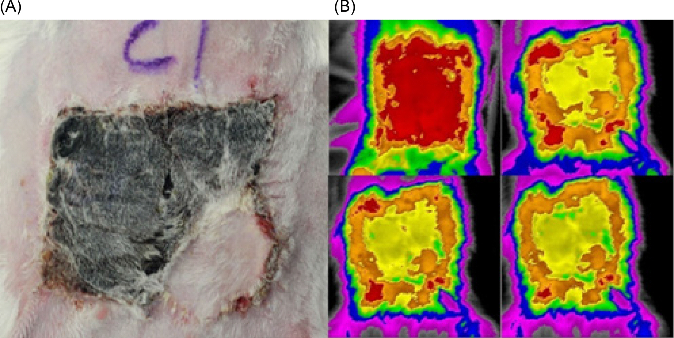
Table 1.Comparison of expected survival area by DITI and measured survival area
Table 2.Statistical analysis by paired T-test
|
Expected survival area by DITI (%) |
Measured survival area (%) |
|
|---|---|---|
| Mean SD | 55.3 (16.6) | 56.2 (18.0) |
| P-value* | 0.6 | 629 |
- 1.Hovius SE, van Adrichem LN, Mulder HD, van Strik R, van der Meulen JC. The predictive value of the laser Doppler flowmeter for postoperative microvascular monitoring. Ann Plast Surg 1993;31:307–12.ArticlePubMed
- 2.Hwang JW, Park DH. Estimation of the normal skin blood flow by laser Doppler flowmetry. J Korean Soc Plast Reconstr Surg 1996;23:394–404.
- 3.Jones BM, Sanders R, Greenhalgh RM. Monitoring skin flaps by colour measurement. Br J Plast Surg 1983;36:88–94.ArticlePubMed
- 4.Khouri RK, Shaw WW. Monitoring of free flaps with surface temperature recordings: is it reliable? Plast Reconstr Surg 1992;89:495–9.PubMed
- 5.Russell JA, Conforti ML, Connor NP, Hartig GK. Cutaneous tissue flap viability following partial venous obstruction. Plast Reconstr Surg 2006;117:2259–66.ArticlePubMed
- 6.van Dam H, Nduka C, Carver N. No touch free-flap temperature monitoring. Br J Plast Surg 2003;56:835.ArticlePubMed
- 7.Lee SH, Baek SH, Hwang WJ, Jo DI, Oh JK, Kim JT. Application of ThermalCAMTM P40-Infrared thermographic imaging in the monitoring of survival of lower abdominal random flap in the rabbit. J Korean Soc Plast Reconstr Surg 2004;31:95–101.
- 8.Rogatto WDThe infrared & electro-optical systems handbook. Electro-optical Components. Vol. 3, Ann Arbor: SPIE Press; 1993. p.177..
- 9.Dunn RM, Mancoll J. Flap models in the rat: a review and reappraisal. Plast Reconstr Surg 1992;90:319–28.ArticlePubMed
- 10.Mirzabeigi MN, Wang T, Kovach SJ, Taylor JA, Serletti JM, Wu LC. Free flap take-back following postoperative microvascular compromise: predicting salvage versus failure. Plast Reconstr Surg 2012;130:579–89.PubMed
- 11.Lee DE, Chung HK, Kim YB, Yang SJ, Park CS. Comparision of assessment of blood flow in flaps with laser Doppler flowmeter and dermatofluorometer. J Korean Soc Plast Reconstr Surg 1995;22:771–80.
- 12.Cha BH, Kim SK, Kim JT. Estimation of lower abdominal flap survival in rabbit model using laser Doppler flowmetry value. J Korean Soc Plast Reconstr Surg 2002;29:311–7.
- 13.Kim YW, Kim MR. Diagnostic efficacy of DITI(Digital Infrared Thermographic Imaging) for the dysesthesia of the lower lip & chin. J Korean Assoc Oral Maxillofac Surg 2002;28:53–60.
- 14.Gratt BM, Sickles EA. Electronic facial thermography: An analysis of asymptomatic adult subjects. J Orofac Pain 1995;9:255–65.PubMed
- 15.Gratt BM, Shetty V, Saiar M, Sickles EA. Electronic thermography for the assessment of inferior alveolar nerve deficit. Oral Surg Oral Med Oral Pathol Oral Radiol Endod 1995;80:153–60.ArticlePubMed
- 16.Biagioni PA, Longmore RB, McGimpsey JG, Lamey PJ. Infrared thermography. Its role in dental research with particular reference to craniomandibular disorders. Dentomaxillofac Radiol 1996;25:119–24.ArticlePubMed
- 17.Kruse RA Jr, Christiansen JA. Thermographic imaging of myofascial trigger points: A follow-up study. Arch Phys Med Rehabil 1992;73:819–23.PubMed
References
Figure & Data
References
Citations
Citations to this article as recorded by 
 PubReader
 ePub Link
 Cite
Cite


