 KOSIN UNIVERSITY COLLEGE OF MEDICINE
Articles
- Page Path
- HOME > Kosin Med J > Volume 38(3); 2023 > Article
-
Review article
Application of Raman spectroscopy in breast cancer surgery -
Yikeun Kim1
 , Sung Ui Jung2, Jinhyuk Choi2
 -
Kosin Medical Journal 2023;38(3):176-183.
DOI: https://doi.org/10.7180/kmj.23.129
Published online: September 14, 2023
1Department of Biomedical Engineering, Ulsan National Institute of Science and Technology (UNIST), Ulsan, Korea
2Department of Surgery, Kosin University Gospel Hospital, Kosin University College of Medicine, Busan, Korea
- Corresponding Author: Jin Hyuk Choi, MD Department of Surgery, Kosin University Gospel Hospital, Kosin University College of Medicine, 262 Gamcheon-ro, Seo-gu, Busan 49267, Korea Tel: +82-51-990-6781 Fax: +82-51-990-6462 E-mail: drchoijinhyuk@gmail.com
Copyright © 2023 Kosin University College of Medicine.
This is an open-access article distributed under the terms of the Creative Commons Attribution Non-Commercial License (http://creativecommons.org/licenses/by-nc/4.0/) which permits unrestricted non-commercial use, distribution, and reproduction in any medium, provided the original work is properly cited.
- 1,337 Views
- 32 Download
Abstract
- The incidence of breast cancer is increasing worldwide. As cancer screening has become more widespread, the rate of early breast cancer detection has increased and treatment methods have changed. Partial mastectomy is performed more often than total mastectomy for the surgical treatment of early breast cancer, and sentinel lymph node biopsy plays an important role. A high level of accuracy is necessary for the intraoperative examination of surgical margins and sentinel lymph nodes to identify malignancies. Therefore, several examination techniques, including Raman spectroscopy, that replace or supplement the currently used frozen-section methods are being studied. Raman spectroscopy has the ability to diagnose cancer in normal tissue by providing in real time a chemical fingerprint that can be used to differentiate between cells and tissues. Numerous studies have investigated the utilization of Raman spectroscopy to identify cancer in the margins of resected tissues and sentinel lymph nodes during breast cancer surgery, showing the potential of this technique for clinical applications. This article introduces and reviews the research on Raman spectroscopy for breast cancer surgery.
- Breast cancer is currently the most commonly diagnosed cancer in women, accounting for a quarter of all cancer cases [1]. A total of 2.3 million new cases of breast cancer were diagnosed in 2020, accounting for one in eight newly diagnosed cancers [2]. The incidence of breast cancer is rising not only in the United States and Europe but also in Asia [3]. With the development of screening using mammography and ultrasound, and the development of various treatment methods, the treatment results for breast cancer are becoming more outstanding [4]. Among all breast cancer diagnoses, the proportion of early-stage breast cancers continues to increase. Accordingly, the surgical methods for breast cancer are constantly changing. Modified radical mastectomy was the standard for surgical treatment of breast cancer. However, with the introduction of partial mastectomy accompanied by radiation, the surgical treatment of breast cancer has undergone great changes [5,6]. In addition, with the introduction of sentinel lymph node biopsy (SLNB) into breast cancer surgery by Giuliano et al. [7], many patients with early breast cancer skipped axillary lymphatic dissection, thereby reducing the incidence of lymphedema. Such changes in breast cancer surgery require higher levels of accuracy and safety. In the process of confirming the resection margin and sentinel lymph nodes by frozen section examination during surgery, a certain false-negative rate is reported in the pathological examination [8,9]. If the frozen biopsy result is negative, but cancer cells are found in the final biopsy report, the patient may experience the inconvenience of having to undergo reoperation to remove any cancer cells that may remain in the body. The reoperation rate has been reported up to 50% depending on the study [10-12]. It follows that optical technologies have been developed for accurate diagnosis of sentinel lymph node and tumor margins such as diffuse reflectance spectroscopy, fluorescence spectroscopy, and photoacoustic spectroscopy [13-18]. Especially Raman spectroscopy is an excellent technique for material analysis due to its high molecular specificity [19], various studies have been conducted on the evaluation of sentinel lymph node and tumor margin in breast cancer [20-24]. But Raman scattering has disadvantages that are difficult to apply clinically, such as low signal-to-noise ratio and exacerbated by fluorescence interference [25], and long measurement time [26]. Nonetheless, recent advances such as high-efficiency laser sources, low-noise detectors, effective filters, and high-efficiency optics have greatly improved this applicability [27,28].
Introduction
- 1. Raman spectroscopy system
- Raman spectroscopy was first observed experimentally in 1928 [29]; however, because of the rare occurrence of Raman scattering, which only occurs with a probability of 1 in approximately 108, it was difficult to observe [30,31]. Recent advancements in technology have enabled real-time observation using Raman spectroscopy, leading to its widespread application in the clinical field. Raman spectroscopy is a powerful technique for the spectroscopy of vibrations produced by the interacting energy in materials, including cells and tissues. It allows the identification and analysis of the molecular structure, symmetry, electronic environment, and composition of a material, providing a chemical fingerprint that can be used to distinguish between cells and tissues [32-37]. Diseases, particularly cancer, alter the chemical fingerprints of tissues. Raman spectroscopy has the potential to differentiate between diseased and normal tissues. However, it requires a large amount of trained reference data and an accurate analysis model [32,34].
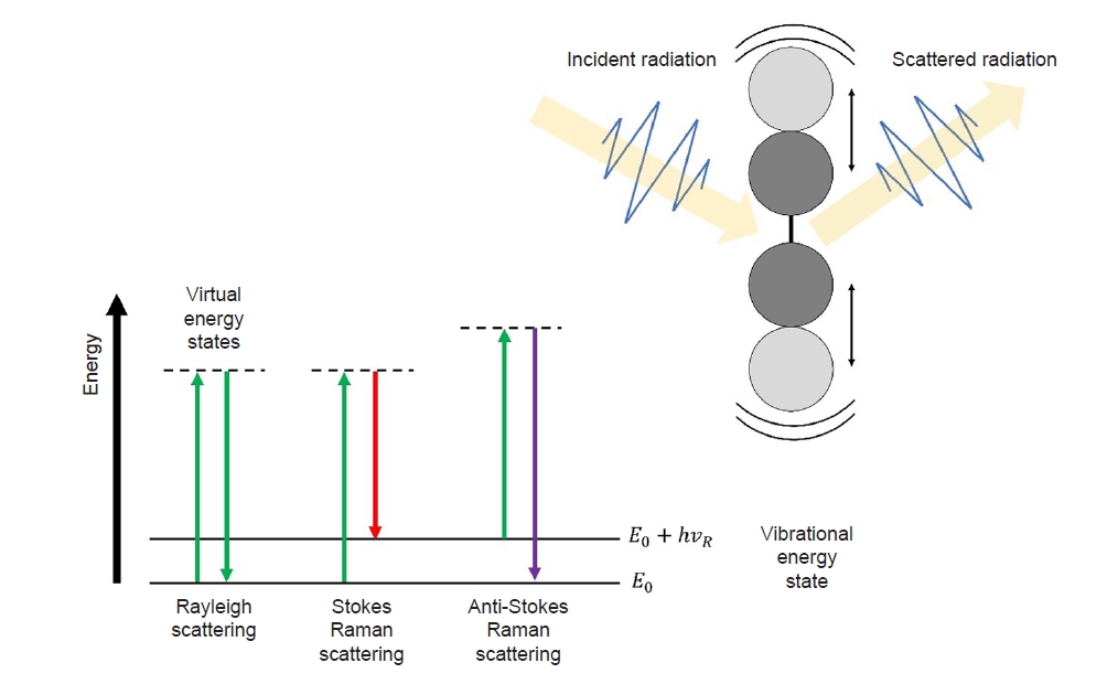
- To explain the Raman scattering phenomenon, it is necessary to first describe Rayleigh scattering. Various interactions, such as absorption, reflection, and scattering, occur when light interacts with a material. Scattering refers to the deviation of light from its original path and its propagation in different directions. Rayleigh scattering occurs when the energies of the incident light and light emitted in different directions are equal. This type of scattering is also known as elastic scattering, owing to its characteristic nature. However, there are cases in which the scattered light possesses more or less energy than the original energy. For example, a portion of the incident energy may be utilized for the vibrational motion of atoms or the rotational motion of molecules, whereas the remaining energy is scattered as light. In this scenario, only energy lower than the incident energy is emitted, resulting in the emission of light with relatively longer wavelengths compared to Rayleigh scattering. This process is referred to as Stokes Raman scattering. Conversely, when the material is already in a high-energy state upon receiving light, more energy is emitted than incident energy. Consequently, the wavelength of the scattered light shortens; this phenomenon is known as anti-Stokes Raman scattering (Fig. 1) [38].
- 2. Raman spectra
- The incident photons interact with molecules in the tissue. Rayleigh occurs when the energy of the scattered photon is equal to that of the incident photon. The rare occurrence of a difference in energy between a scattered photon and an incident photon is called inelastic scattering and is known as the Raman effect. Only one photon in 108 undergoes the Raman effect [30,39].
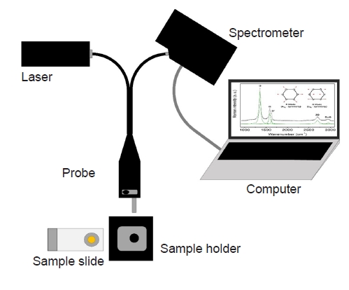
- A laser is used to provide high-quality monochromatic light and induce Raman scattering. To collect light that reacts with the tissue, appropriate optics must be configured in the optical path. Recent developments in Raman detectors include the use of highly sensitive detectors and gratings. A schematic of the Raman spectroscopy system is shown in Fig. 2.
- In general, Raman spectra have characteristic chemical fingerprints that depend on the wavelength of the incident laser, molecular composition, and bonding form. Raman spectroscopic techniques have been developed in several types. Spontaneous Raman spectroscopy, when used in combination with a fiber probe or microscope, is characterized by being label-free, noninvasive, and nondestructive. Resonance Raman spectroscopy, which matches the excitation wavelength to the electron resonance of molecules, increases the signal-to-noise ratio by 103 to 105. Surface-enhanced Raman spectroscopy applied to rough metal surfaces results in a 106-fold increase in the signal-to-noise ratio and has been applied to cell-based assays and immunoassays. Spatially offset Raman spectroscopy technology, which collects diffusely scattered photons, can acquire information even in relatively thick tissues and has been applied to cancer detection in breast tissue [30].
- The Raman spectrum provides the fingerprint of a material; however, it is not possible to directly interpret the composition of the material from this chemical fingerprint. A database of reference spectra is required to use Raman spectroscopy in the analysis of materials. A large number of Raman spectra have been published for this purpose [40-42]. Also, simulations and deep learning continue to be studied [39,43]. Unlike single materials, human tissues are a complex assembly of various molecular structures. Raman spectra of protein structures in human tissues are continuously being studied [44,45].
Background of Raman spectroscopy
- 1. Frozen section analysis in breast cancer surgery
- Partial mastectomy combined with postoperative radiation therapy has become the gold standard treatment for patients with early-stage breast cancer, offering equivalent survival and improved quality of life compared to patients undergoing total mastectomy [46,47]. Complete resection of tumors is essential for partial mastectomy to reduce the recurrence rate after surgical treatment [48,49]. During partial mastectomy, the surgeon may request rapid pathological information of marginal status and determine whether additional resection is required. SLNB is also important for the surgical treatment of early breast cancer. The surgeon checks the progress of the disease through SLNB during surgery and makes a dicision of the surgical scope of the axillary region. As the rate of early breast cancer increases, the need for a method that can quickly confirm pathological results during surgery has increased. Currently, frozen section analysis is the most frequently performed method for confirming the pathological results of intraoperative biopsies. When a surgeon sends a tissue that needs to be inspected during surgery to a pathologist, the tissue is analyzed through frozen sectioning and notified of the result, which reduces the frequency of reoperations that occur after surgery [50,51]. However, This method has limitation in sensitivity. Studies analyzing marginal frozen sections obtained from partial mastectomy have reported sensitivities of 77%–81% [52,53]. In SLNB, the sensitivity of macro-metastasis and micro-metastasis was different. The sensitivity for diagnosis of macro-metastasis was over 90%, but the sensitivity for diagnosis of micro-metastasis was reported to be 30%–40% [8,54-56]. If the results of frozen section analysis confirmed during surgery and the final biopsy results confirmed after surgery are different, the patient may experience the inconvenience of having to repeat the operation, which leads to an increase in complications, hospitalization days, and medical expenses [57-60].
- 2. Raman spectroscopy of surgical margins
- In 2006, a study that implemented Raman spectroscopy of breast tissue in an in vivo environment was reported for the first time. Haka et al. [61] obtained and analyzed 31 Raman spectra from nine patients who underwent partial mastectomy. In that study, cancer tissues were accurately distinguished from normal and benign tissues using Raman spectroscopy. These researchers later reported a negative predictive value of 99% using 129 tissue samples in a new prospective study [62]. However, to confirm the presence or absence of cancerous tissue on the surgical cut surface using Raman spectroscopy in actual clinical practice, an accurate location must be specified, and the single-point method using a probe causes sampling errors. To solve this problem, Zhang et al. [63] conducted a comparative study using Raman spectral mapping. A total of 53 sets of mapping data and 2,597 Raman spectra were analyzed and compared, and the data obtained using the mapping technology displayed excellent diagnostic performance. Raman microspectroscopy studies have also been reported. Raman microspectroscopy makes diagnosis without staining based on the morphological and biochemical contrast between normal and tumor tissue. Kong et al. [64] reported that using Raman microspectroscopy to detect invasive ductal carcinoma within breast tissue with 95.6% sensitivity and 96.2% specificity. Zhang et al. [65] reported characterization of biochemical properties and structural alterations of breast cancer tissues at various TNM stages and grades by Raman microspectroscopy. Early Raman microspectroscopy studies had limitations in that the scanning method used to construct Raman spectral images for tumor diagnosis was very slow [66]. However, with the development of various technologies, such as the use of selective sampling based on integrated autofluorescence imaging, the possibility of its clinical application as an intraoperative method has been demonstrated [64,67,68].
- 3. Raman spectroscopy of SLNB
- Raman spectroscopy is noninvasive and can provide detailed chemical information about tissue, thereby making it a very suitable test to check the status of the sentinel lymph nodes in real time during surgery. In 2003, Smith et al. [69] first identified axillary lymph nodes in breast cancer using Raman spectroscopy. After that, Horsnell et al. [70] reported a sensitivity of 81% and specificity of 97% using the method of examining 10 points in the lymph node. Unfortunately, studies using Raman spectroscopy as a diagnostic tool for sentinel lymph node evaluation have not yet been conducted. Most studies were limited to small sample sizes and were laboratory-based. However, recently, studies using new technologies, such as a tissue mapping protocol obtained by analyzing the spectra of each cell [22] and studies using a nontoxic Raman nanoparticle tracer [21] have been reported, confirming the possibility that sentinel lymph node diagnosis through Raman spectroscopy can be used in clinical practice.
Raman spectroscopy of breast cancer surgery
- Research on Raman spectroscopy has been conducted in various fields, ranging from basic to clinical applications. The high sensitivity of Raman spectroscopy was previously regarded as a disadvantage that made it difficult to apply in clinical practice; however, these limitations are now being overcome by the incorporation of various technologies and the development of spectrum analysis. Studies analyzing Raman spectra to identify cancerous tissue at the surgical margin and lymph node during breast cancer surgery are ongoing, and the positive results of the studies show the possibility of supplementing the frozen section method. In the surgical treatment of breast cancer, if it becomes possible to distinguish malignant tissue from normal tissue in vivo using Raman spectroscopy, unnecessary surgical biopsies during surgery will be reduced.
Conclusions
-
Conflicts of interest
No potential conflict of interest relevant to this article was reported.
-
Funding
This work was supported by the Basic Science Research Program through the National Research Foundation of Korea (NRF), funded by the Ministry of Education (NRF-2021R1G1A1011865).
-
Author contributions
Conceptualization: YK, JC. Funding acquisition: JC. Writing - original draft: YK, SUJ. Writing - review & editing: YK, SUJ, JC.
Article information
- 1. Heer E, Harper A, Escandor N, Sung H, McCormack V, Fidler-Benaoudia MM. Global burden and trends in premenopausal and postmenopausal breast cancer: a population-based study. Lancet Glob Health 2020;8:e1027–37.ArticlePubMed
- 2. Sung H, Ferlay J, Siegel RL, Laversanne M, Soerjomataram I, Jemal A, et al. Global cancer statistics 2020: GLOBOCAN estimates of incidence and mortality worldwide for 36 cancers in 185 countries. CA Cancer J Clin 2021;71:209–49.ArticlePubMedPDF
- 3. Lei S, Zheng R, Zhang S, Wang S, Chen R, Sun K, et al. Global patterns of breast cancer incidence and mortality: a population-based cancer registry data analysis from 2000 to 2020. Cancer Commun (Lond) 2021;41:1183–94.ArticlePubMedPMCPDF
- 4. Arnold M, Morgan E, Rumgay H, Mafra A, Singh D, Laversanne M, et al. Current and future burden of breast cancer: global statistics for 2020 and 2040. Breast 2022;66:15–23.ArticlePubMedPMC
- 5. Early Breast Cancer Trialists’ Collaborative Group. Effects of radiotherapy and surgery in early breast cancer: an overview of the randomized trials. N Engl J Med 1995;333:1444–56.ArticlePubMed
- 6. Kim YS, Ryu DW, Lee CH. Comparison of survival outcomes between modified radical mastectomy and breast conserving surgery in early breast cancer patients. Kosin Med J 2016;31:19–29.ArticlePDF
- 7. Giuliano AE, Jones RC, Brennan M, Statman R. Sentinel lymphadenectomy in breast cancer. J Clin Oncol 1997;15:2345–50.ArticlePubMed
- 8. Elshanbary AA, Awad AA, Abdelsalam A, Ibrahim IH, Abdel-Aziz W, Darwish YB, et al. The diagnostic accuracy of intraoperative frozen section biopsy for diagnosis of sentinel lymph node metastasis in breast cancer patients: a meta-analysis. Environ Sci Pollut Res Int 2022;29:47931–41.ArticlePubMedPMCPDF
- 9. Holck S, Galatius H, Engel U, Wagner F, Hoffmann J. False-negative frozen section of sentinel lymph node biopsy for breast cancer. Breast 2004;13:42–8.ArticlePubMed
- 10. McLaughlin SA, Ochoa-Frongia LM, Patil SM, Cody HS 3rd, Sclafani LM. Influence of frozen-section analysis of sentinel lymph node and lumpectomy margin status on reoperation rates in patients undergoing breast-conservation therapy. J Am Coll Surg 2008;206:76–82.ArticlePubMed
- 11. Aziz D, Rawlinson E, Narod SA, Sun P, Lickley HL, McCready DR, et al. The role of reexcision for positive margins in optimizing local disease control after breast-conserving surgery for cancer. Breast J 2006;12:331–7.ArticlePubMed
- 12. Jacobson AF, Asad J, Boolbol SK, Osborne MP, Boachie-Adjei K, Feldman SM. Do additional shaved margins at the time of lumpectomy eliminate the need for re-excision? Am J Surg 2008;196:556–8.ArticlePubMed
- 13. Wilke LG, Brown JQ, Bydlon TM, Kennedy SA, Richards LM, Junker MK, et al. Rapid noninvasive optical imaging of tissue composition in breast tumor margins. Am J Surg 2009;198:566–74.ArticlePubMedPMC
- 14. Kennedy S, Geradts J, Bydlon T, Brown JQ, Gallagher J, Junker M, et al. Optical breast cancer margin assessment: an observational study of the effects of tissue heterogeneity on optical contrast. Breast Cancer Res 2010;12:R91.ArticlePubMedPMCPDF
- 15. Tellier F, Ravelo R, Simon H, Chabrier R, Steibel J, Poulet P. Sentinel lymph node detection by an optical method using scattered photons. Biomed Opt Express 2010;1:902–10.ArticlePubMedPMC
- 16. Lu Y, Zhao Y, Zhu Y, Xu X, Yin J. In situ research and diagnosis of breast cancer by using HOF-ATR-FTIR spectroscopy. Spectrochim Acta A Mol Biomol Spectrosc 2020;235:118178.ArticlePubMed
- 17. Phipps JE, Gorpas D, Unger J, Darrow M, Bold RJ, Marcu L. Automated detection of breast cancer in resected specimens with fluorescence lifetime imaging. Phys Med Biol 2017;63:015003.ArticlePubMedPMCPDF
- 18. Balasundaram G, Goh Y, Moothanchery M, Attia A, Lim HQ, Burton NC, et al. Optoacoustic characterization of breast conserving surgery specimens: a pilot study. Photoacoustics 2020;19:100164.ArticlePubMedPMC
- 19. Butler HJ, Ashton L, Bird B, Cinque G, Curtis K, Dorney J, et al. Using Raman spectroscopy to characterize biological materials. Nat Protoc 2016;11:664–87.ArticlePubMedPDF
- 20. Bao Z, Zhang Y, Tan Z, Yin X, Di W, Ye J. Gap-enhanced Raman tags for high-contrast sentinel lymph node imaging. Biomaterials 2018;163:105–15.ArticlePubMed
- 21. Deng B, Wang Y, Wu Y, Yin W, Lu J, Ye J. Raman nanotags-guided intraoperative sentinel lymph nodes precise location with minimal invasion. Adv Sci (Weinh) 2022;9:e2102405.ArticlePubMedPDF
- 22. Som D, Tak M, Setia M, Patil A, Sengupta A, Chilakapati CM, et al. A grid matrix-based Raman spectroscopic method to characterize different cell milieu in biopsied axillary sentinel lymph nodes of breast cancer patients. Lasers Med Sci 2016;31:95–111.ArticlePubMedPDF
- 23. Zuniga WC, Jones V, Anderson SM, Echevarria A, Miller NL, Stashko C, et al. Raman spectroscopy for rapid evaluation of surgical margins during breast cancer lumpectomy. Sci Rep 2019;9:14639.ArticlePubMedPMCPDF
- 24. Keller MD, Vargis E, de Matos Granja N, Wilson RH, Mycek MA, Kelley MC, et al. Development of a spatially offset Raman spectroscopy probe for breast tumor surgical margin evaluation. J Biomed Opt 2011;16:077006.ArticlePubMedPMC
- 25. Vankeirsbilck T, Vercauteren A, Baeyens W, Van der Weken G, Verpoort F, Vergote G, et al. Applications of Raman spectroscopy in pharmaceutical analysis. TrAC Trends Anal Chem 2002;21:869–77.Article
- 26. Eberhardt K, Stiebing C, Matthaus C, Schmitt M, Popp J. Advantages and limitations of Raman spectroscopy for molecular diagnostics: an update. Expert Rev Mol Diagn 2015;15:773–87.ArticlePubMed
- 27. Zhou J, Pan W, Qi W, Cao X, Cheng Z, Feng Y. Ultrafast Raman fiber laser: a review and prospect. PhotoniX 2022;3:18.ArticlePDF
- 28. Ye J, Ma X, Zhang Y, Xu J, Zhang H, Yao T, et al. From spectral broadening to recompression: dynamics of incoherent optical waves propagating in the fiber. PhotoniX 2021;2:15.ArticlePDF
- 29. Raman CV, Krishnan KS. A new type of secondary radiation. Nature 1928;121:501–2.ArticlePDF
- 30. Ember KJ, Hoeve MA, McAughtrie SL, Bergholt MS, Dwyer BJ, Stevens MM, et al. Raman spectroscopy and regenerative medicine: a review. NPJ Regen Med 2017;2:12.ArticlePubMedPMCPDF
- 31. Choo-Smith LP, Edwards HG, Endtz HP, Kros JM, Heule F, Barr H, et al. Medical applications of Raman spectroscopy: from proof of principle to clinical implementation. Biopolymers 2002;67:1–9.ArticlePubMed
- 32. Kong K, Kendall C, Stone N, Notingher I. Raman spectroscopy for medical diagnostics: from in-vitro biofluid assays to in-vivo cancer detection. Adv Drug Deliv Rev 2015;89:121–34.ArticlePubMed
- 33. Hanlon EB, Manoharan R, Koo TW, Shafer KE, Motz JT, Fitzmaurice M, et al. Prospects for in vivo Raman spectroscopy. Phys Med Biol 2000;45:R1–59.ArticlePubMed
- 34. Kallaway C, Almond LM, Barr H, Wood J, Hutchings J, Kendall C, et al. Advances in the clinical application of Raman spectroscopy for cancer diagnostics. Photodiagnosis Photodyn Ther 2013;10:207–19.ArticlePubMed
- 35. Das RS, Agrawal YK. Raman spectroscopy: recent advancements, techniques and applications. Vib Spectrosc 2011;57:163–76.Article
- 36. Vaskova H. A powerful tool for material identification: Raman spectroscopy. Int J Math Model Methods Appl Sci 2011;5:1205–12.
- 37. Jones RR, Hooper DC, Zhang L, Wolverson D, Valev VK. Raman techniques: fundamentals and frontiers. Nanoscale Res Lett 2019;14:231.ArticlePubMedPMCPDF
- 38. Shipp DW, Sinjab F, Notingher I. Raman spectroscopy: techniques and applications in the life sciences. Adv Opt Photonics 2017;9:315–428.Article
- 39. Bagheri M, Komsa HP. High-throughput computation of Raman spectra from first principles. Sci Data 2023;10:80.ArticlePubMedPMCPDF
- 40. El Mendili Y, Vaitkus A, Merkys A, Grazulis S, Chateigner D, Mathevet F, et al. Raman Open Database: first interconnected Raman-X-ray diffraction open-access resource for material identification. J Appl Crystallogr 2019;52(Pt 3):618–25.ArticlePubMedPMC
- 41. Grazulis S, Chateigner D, Downs RT, Yokochi AF, Quiros M, Lutterotti L, et al. Crystallography Open Database: an open-access collection of crystal structures. J Appl Crystallogr 2009;42(Pt 4):726–9.ArticlePubMedPMC
- 42. Grazulis S, Daskevic A, Merkys A, Chateigner D, Lutterotti L, Quiros M, et al. Crystallography Open Database (COD): an open-access collection of crystal structures and platform for world-wide collaboration. Nucleic Acids Res 2012;40(Database issue):D420–7.ArticlePubMed
- 43. Huang L, Sun H, Sun L, Shi K, Chen Y, Ren X, et al. Rapid, label-free histopathological diagnosis of liver cancer based on Raman spectroscopy and deep learning. Nat Commun 2023;14:48.ArticlePubMedPMCPDF
- 44. Fujita K, Smith NI. Label-free molecular imaging of living cells. Mol Cells 2008;26:530–5.ArticlePubMed
- 45. Xu J, Yu T, Zois CE, Cheng JX, Tang Y, Harris AL, et al. Unveiling cancer metabolism through spontaneous and coherent Raman spectroscopy and stable isotope probing. Cancers (Basel) 2021;13:1718.ArticlePubMedPMC
- 46. Fisher B, Anderson S, Bryant J, Margolese RG, Deutsch M, Fisher ER, et al. Twenty-year follow-up of a randomized trial comparing total mastectomy, lumpectomy, and lumpectomy plus irradiation for the treatment of invasive breast cancer. N Engl J Med 2002;347:1233–41.ArticlePubMed
- 47. Veronesi U, Cascinelli N, Mariani L, Greco M, Saccozzi R, Luini A, et al. Twenty-year follow-up of a randomized study comparing breast-conserving surgery with radical mastectomy for early breast cancer. N Engl J Med 2002;347:1227–32.ArticlePubMed
- 48. Obedian E, Haffty BG. Negative margin status improves local control in conservatively managed breast cancer patients. Cancer J Sci Am 2000;6:28–33.PubMed
- 49. Yang SI, Lee SH. Clinical outcome of positive margin of postgastrectomy with adenocarcinoma of stomach. Kosin Med J 2012;27:31–6.ArticlePDF
- 50. Farouk O, Senbel A, Shetiwy M, Attia E, Abdallah A, El-Damshety O, et al. The effectiveness of intraoperative frozen section analysis of safety margins in breast conserving surgery and the role of surgeon in decreasing the rate of positive margins. Surg Sci 2017;8:499–509.
- 51. Nowikiewicz T, Srutek E, Glowacka-Mrotek I, Tarkowska M, Zyromska A, Zegarski W. Clinical outcomes of an intraoperative surgical margin assessment using the fresh frozen section method in patients with invasive breast cancer undergoing breast-conserving surgery: a single center analysis. Sci Rep 2019;9:13441.ArticlePubMedPMCPDF
- 52. Noguchi M, Minami M, Earashi M, Taniya T, Miyazaki I, Mizukami Y, et al. Intraoperative histologic assessment of surgical margins and lymph node metastasis in breast-conserving surgery. J Surg Oncol 1995;60:185–90.ArticlePubMed
- 53. Garcia MT, Mota BS, Cardoso N, Martimbianco AL, Ricci MD, Carvalho FM, et al. Accuracy of frozen section in intraoperative margin assessment for breast-conserving surgery: a systematic review and meta-analysis. PLoS One 2021;16:e0248768.ArticlePubMedPMC
- 54. Schrenk P, Wayand W. Sentinel-node biopsy in axillary lymph-node staging for patients with multicentric breast cancer. Lancet 2001;357:122.ArticlePubMed
- 55. Morgan A, Howisey RL, Aldape HC, Patton RG, Rowbotham RK, Schmidt EK, et al. Initial experience in a community hospital with sentinel lymph node mapping and biopsy for evaluation of axillary lymph node status in palpable invasive breast cancer. J Surg Oncol 1999;72:24–30.ArticlePubMed
- 56. Weiser MR, Montgomery LL, Susnik B, Tan LK, Borgen PI, Cody HS. Is routine intraoperative frozen-section examination of sentinel lymph nodes in breast cancer worthwhile? Ann Surg Oncol 2000;7:651–5.ArticlePubMedPDF
- 57. McCahill LE, Single RM, Aiello Bowles EJ, Feigelson HS, James TA, Barney T, et al. Variability in reexcision following breast conservation surgery. JAMA 2012;307:467–75.ArticlePubMed
- 58. Jorns JM, Daignault S, Sabel MS, Wu AJ. Is intraoperative frozen section analysis of reexcision specimens of value in preventing reoperation in breast-conserving therapy? Am J Clin Pathol 2014;142:601–8.ArticlePubMed
- 59. Jorns JM, Visscher D, Sabel M, Breslin T, Healy P, Daignaut S, et al. Intraoperative frozen section analysis of margins in breast conserving surgery significantly decreases reoperative rates: one-year experience at an ambulatory surgical center. Am J Clin Pathol 2012;138:657–69.ArticlePubMed
- 60. Lessells AM, Simpson JG. A retrospective analysis of the accuracy of immediate frozen section diagnosis in surgical pathology. Br J Surg 1976;63:327–9.ArticlePubMedPDF
- 61. Haka AS, Volynskaya Z, Gardecki JA, Nazemi J, Lyons J, Hicks D, et al. In vivo margin assessment during partial mastectomy breast surgery using Raman spectroscopy. Cancer Res 2006;66:3317–22.ArticlePubMedPDF
- 62. Haka AS, Volynskaya Z, Gardecki JA, Nazemi J, Shenk R, Wang N, et al. Diagnosing breast cancer using Raman spectroscopy: prospective analysis. J Biomed Opt 2009;14:054023.ArticlePubMedPMC
- 63. Zhang H, Wang X, Ding R, Shen L, Gao P, Xu H, et al. Characterization and imaging of surgical specimens of invasive breast cancer and normal breast tissues with the application of Raman spectral mapping: a feasibility study and comparison with randomized single-point detection method. Oncol Lett 2020;20:2969–76.ArticlePubMedPMC
- 64. Kong K, Zaabar F, Rakha E, Ellis I, Koloydenko A, Notingher I. Towards intra-operative diagnosis of tumours during breast conserving surgery by selective-sampling Raman micro-spectroscopy. Phys Med Biol 2014;59:6141–52.ArticlePubMedPDF
- 65. Zhang B, Zhang Z, Gao B, Zhang F, Tian L, Zeng H, et al. Raman microspectroscopy based TNM staging and grading of breast cancer. Spectrochim Acta A Mol Biomol Spectrosc 2023;285:121937.ArticlePubMed
- 66. Kong K, Rowlands CJ, Varma S, Perkins W, Leach IH, Koloydenko AA, et al. Diagnosis of tumors during tissue-conserving surgery with integrated autofluorescence and Raman scattering microscopy. Proc Natl Acad Sci U S A 2013;110:15189–94.ArticlePubMedPMC
- 67. Horsnell JD, Kendall C, Stone N. Towards the intra-operative use of Raman spectroscopy in breast cancer-overcoming the effects of theatre lighting. Lasers Med Sci 2016;31:1143–9.ArticlePubMedPDF
- 68. Hanna K, Krzoska E, Shaaban AM, Muirhead D, Abu-Eid R, Speirs V. Raman spectroscopy: current applications in breast cancer diagnosis, challenges and future prospects. Br J Cancer 2022;126:1125–39.ArticlePubMedPDF
- 69. Smith J, Kendall C, Sammon A, Christie-Brown J, Stone N. Raman spectral mapping in the assessment of axillary lymph nodes in breast cancer. Technol Cancer Res Treat 2003;2:327–32.ArticlePubMedPDF
- 70. Horsnell JD, Smith JA, Sattlecker M, Sammon A, Christie-Brown J, Kendall C, et al. Raman spectroscopy: a potential new method for the intra-operative assessment of axillary lymph nodes. Surgeon 2012;10:123–7.ArticlePubMed
 PubReader
 ePub Link
 Cite
Cite

